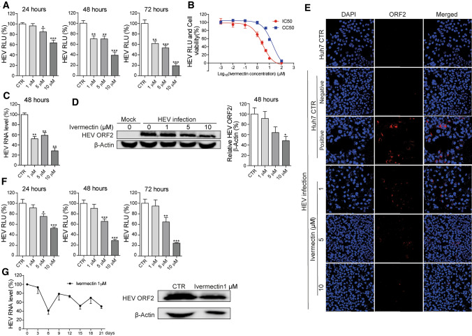
Fig. 1.
Anti-HEV activity of ivermectin in Huh7-based cell culture models. (A) Effect of ivermectin treatment for 24, 48, or 72 hours on viral replication-dependent luciferase activity in the genotype 3 subgenomic Huh7-p6-Luc cell model. The untreated group served as a control (CTR) (set as 100%) (n = 12). (B) The 50% inhibitory concentration (IC50) and 50% cytotoxic concentration (CC50) of ivermectin in Huh7-p6-Luc cell model and Huh7 cell line were calculated using GraphPad Prism 5 software (n = 6-12). (C) The infectious Huh7-p6 cell model was treated with the indicated concentrations of ivermectin for 48 hours. The effect on viral RNA production was quantified by qRT-PCR using primers targeting the ORF2/ORF3 overlap region (n = 4-8). (D) Western blot analysis of the HEV capsid protein level in Huh7-p6 cells treated with ivermectin for 48 hours. The uninfected group (mock) served as a negative control, and the infected but untreated group served as a positive control (set as 100%) (n = 4). (E) Immunofluorescence analysis of viral ORF2-encoded capsid protein (red) in Huh7 cells treated with the indicated concentrations of ivermectin for 48 hours. Huh7 cells incubated with an anti-HEV capsid protein antibody or Huh7-p6 cells incubated with the matched IgG control antibody served as a negative control. Untreated HEV-infected Huh7 cells incubated with the anti-HEV capsid protein antibody served as a positive control. DAPI (blue) was applied to visualize nuclei (40 × oil immersion objective). (F) Huh7 cells containing the genotype 1 HEV replicon (Sar55 clone) were treated with ivermectin for 24, 48, or 72 hours, and viral-replication-dependent luciferase activity was measured (n = 10). (G) The effects of long-term treatment with 1 μM ivermectin on HEV RNA replication in the Huh7-p6 cell model were analyzed by qRT-PCR. The untreated (CTR) group served as a control (set as 100%) (n = 3-4). Western blot of the HEV ORF2-encoded capsid protein performed after 21 days of treatment. RLU, relative luciferase units. Data are presented as the mean ± SEM (*, P < 0.05; **, P < 0.01; ***, P < 0.001).

