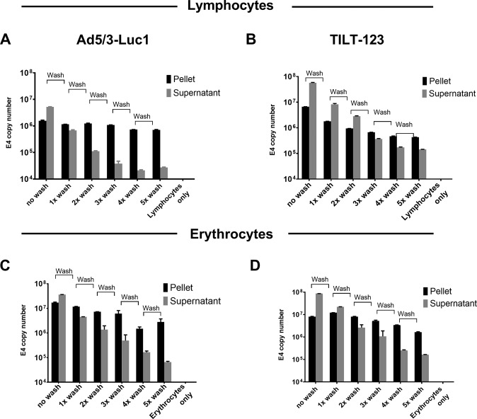
Fig. 1. Association of adenovirus particles with lymphocytes and erythrocytes.
Adenovirus Ad5/3-Luc1 (a, c) and TILT-123 (b, d) were incubated with human lymphocytes at 10 VP/cell (3 × 10e7 VP/ml with 3 × 10e6 cells/ml) (a, b) and with erythrocytes at 0.036 VP/cell (5 × 10e9 VP/ml and 1.8 × 10e8 cells/ml) (c, d) at 37°C. After 30 min incubation, cellular fraction and supernatant were isolated through centrifugation at 2000 × g for 10 min. Cellular fraction was washed five times and after each wash a sample from the supernatant and pellet was collected and analyzed through qPCR. Viral copy number was normalized against amount of genomic DNA in the sample, determined by the expression level of human β-actin. Data are presented as mean + SEM. Adenovirus particles associated with cell fraction (black bar) and supernatant (Sup, gray bars). No wash; first centrifugation after incubation, 1x wash; washed with PBS once, 2x wash; washed twice, 3x wash; 4x wash; 5x wash; washed three, four and five times, respectively.

