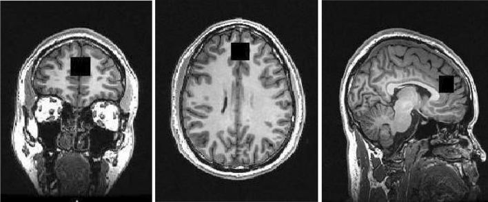
Fig. 1.
Coronal (left), axial (middle), and sagittal (right) planes depicting the placement of the voxel over the anterior cingulate during the proton magnetic resonance spectroscopy scan. The placement of the anterior cingulate voxel was based on the midline sagittal localizer with the centre of the 20 mm × 20 mm × 20 mm voxel placed 13 mm above the anterior portion of the genu of the corpus callosum, perpendicular to the anterior commissure–posterior commissure line to minimize the inclusion of white matter and cerebral spinal fluid

