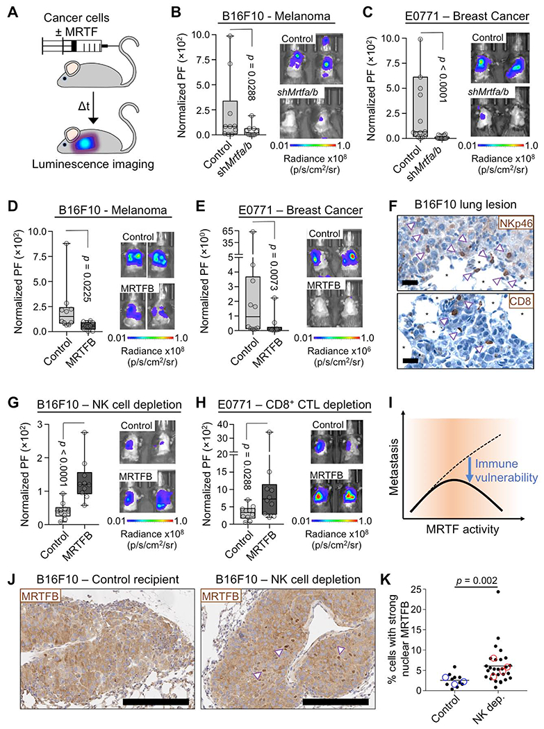
Fig. 1. MRTF overexpression sensitizes metastatic tumors to cytotoxic lymphocytes.

(A) Experimental design for the lung colonization model. (B-C) Metastatic burden in lungs of C57BL/6J mice injected with syngeneic control or Mrtfa/bRNAi B16F10 (B) or E0771 (C) cells, measured by bioluminescent imaging (BLI) 3 weeks after tail vein injection and normalized to the first day of injection. PF: photon flux (n = 10 mice per group). (D-E) BLI of mice 3 weeks post tail vein injection with B16F10 (D) or E0771 (E) cells overexpressing MRTFB or empty vector control (n = 10 mice per group). (F) Representative IHC images of NK cells (arrowheads, NKp46 staining) and CD8+ T cells (arrowheads, CD8 staining) in B16F10 lung metastases. *: alveolar space, scale bars: 20 μm. (G-H) BLI of mice pretreated with anti-asialo GM1 antibody (G) or anti-CD8 antibody (H) for NK and CD8+ T cell depletion, respectively, and imaged 2 weeks after injection of B16F10 (G) or E0771 (H) cells overexpressing MRTFB or empty vector control (n = 10 mice per group). (I) Increased MRTF activity promotes metastasis by enhancing invasiveness (dashed line), but it also sensitizes metastatic cells to cytotoxic lymphocytes (solid line). (J-K) B16F10 lung metastases from control and anti-asialo GM1 treated (NK cell depletion) mice were stained for MRTFB. (J) Representative IHC images, with arrowheads indicating examples of strong nuclear localization. Scale bars: 200 μm. (K) Percentage of cells in each type of tumor with MRTFB nuclear/cytoplasmic ratio > 2 (strong nuclear MRTFB). Closed symbols represent individual lesions (n ≥ 10 lesions of > 200 cells per group), and open symbols denote individual mice (n ≥ 3 per group). Statistical significance was evaluated using lesion data. All p values calculated by Mann-Whitney test. See also Fig. S1 and S2.
