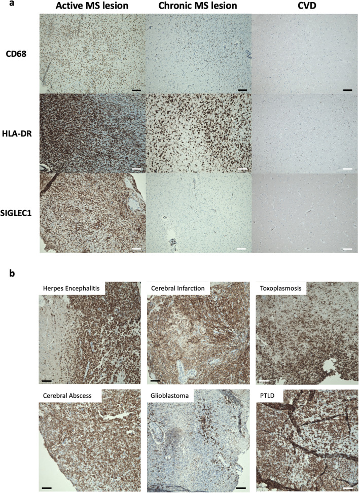
Figure 2.
(a) Representative immunohistochemical stainings of CD68, HLA-DR and SIGLEC1 (CD169) in brain tissue from a patient with active inflammatory multiple sclerosis lesion, a chronic lesion from a patient with secondary-progressive multiple sclerosis (SPMS) and a control patient who died of cardiovascular disease (CVD). Both RRMS and SPMS show infiltrates with CD68+ and HLA-DR+ myeloid cells, but only in the active lesion, these express SIGLEC1. Nuclei are stained blue and the respective marker antigens in brown. The scale bar indicates 100 µm. These results are representative of 4 RRMS, 5 SPMS and 7 control samples that were examined. (b) Immunohistochemical stainings of SIGLEC1 in brain tissue from patients with Herpes simplex viral encephalitis (n = 2), cerebral infarction with inflammatory changes (n = 2), cerebral toxoplasmosis (n = 1), cerebral abscess (n = 2), glioblastoma (n = 2) and post-transplant lymphoproliferative disease (PTLD, n = 1). Infiltrates of SIGLEC1+ myeloid cells were observed in all samples.

