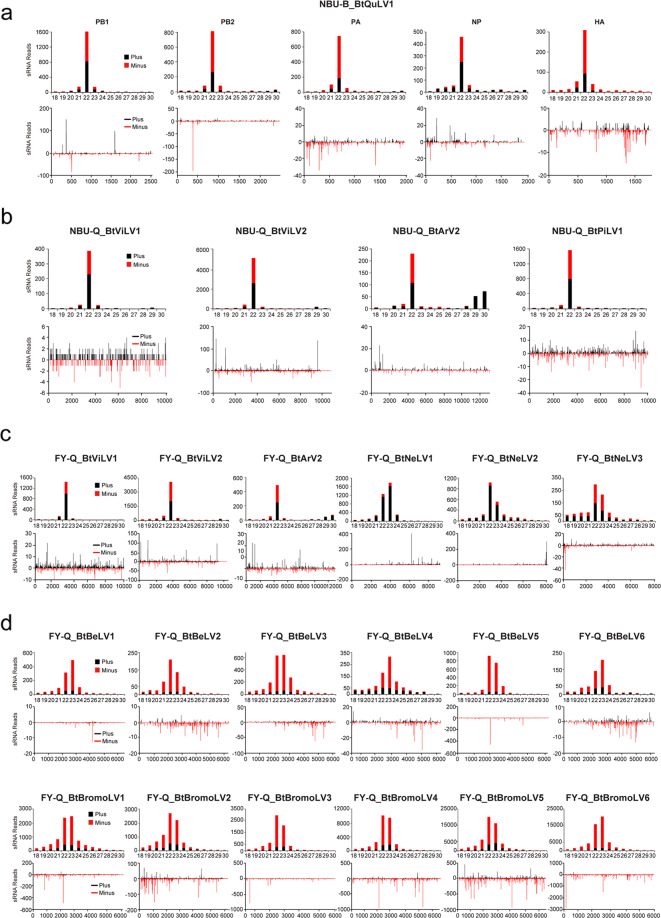
Fig. 5. Profiles of virus-derived small interfering RNAs (vsiRNAs).
vsiRNAs derived from whitefly datasets NBU-B (a), NBU-Q (b), and FY-Q (c, d). The upper panel shows the size distribution of vsiRNAs, while the lower panel shows the distribution of vsiRNAs along the corresponding viral genome. Color coding shows vsiRNAs derived from the sense (black, plus) and antisense (red, minus) genomic strands. All reads in this analysis are redundant. The abbreviation of the virus names is listed in Table 2.

