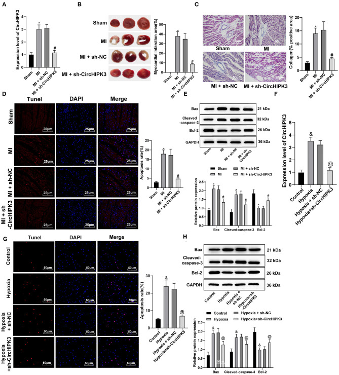
Figure 2.
CircHIPK3 downregulation can reduce cardiomyocyte apoptosis after myocardial infarction (MI) and improve cardiac function. (A) RT-qPCR detected the expression of CircHIPK3 in myocardial tissue. (B) 2,3,5-Triphenyltetrazolium chloride staining was used to detect infarct size. (C) Masson staining was used to detect collagen deposition. (D) TUNEL staining was used to observe the apoptosis of myocardial tissue. (E) The expression of Bax, cleaved caspase-3, and Bcl-2 protein, respectively, was detected by Western blot analysis. (F) RT-qPCR detected CircHIPK3 expression in cardiomyocytes in vitro. (G) The apoptosis of cardiomyocytes was detected by TUNEL staining. (H) Western blot analysis detected the expression of Bax, cleaved caspase-3, and Bcl-2 protein. The value in the figures is the measurement data, which is expressed as mean ± standard deviation. The comparison among multiple groups was analyzed by one-way ANOVA, followed by Tukey's multiple-comparisons test. *compared with sham group, p < 0.05; #compared with MI + sh-NC group, p < 0.05; &compared with control group, p < 0.05; @compared with hypoxia + sh-NC group, p < 0.05. The experiment was repeated three times.

