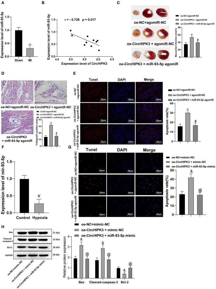
Figure 4.
Overexpression of miR-93-5p reverses the myocardial infarction-induced myocardial injury stimulated by CircHIPK3. (A) RT-PCR was used to detect the expression of miR-93-5p in myocardial tissue of mice after modeling. (B) Pearson's correlation analysis was used to analyze the correlation between miR-93-5p and CircHIPK3. (C) 2,3,5-Triphenyltetrazolium chloride staining was used to detect infarct size. (D) Masson staining was used to detect collagen deposition. (E) TUNEL staining was used to observe the apoptosis of myocardial tissue, n = 10. (F) RT-qPCR detected miR-93-5p expression in cardiomyocytes in vitro. (G) The apoptosis of cardiomyocytes was detected by TUNEL staining. (H) Western blot analysis detected the expression of Bax, cleaved caspase-3, and Bcl-2 protein. The value in the figures is the measurement data, which is expressed as mean ± standard deviation. The comparison between two groups was analyzed by independent-sample t-test. The correlation of circHIPK3 and miR-93-5p was analyzed using Pearson's correlation analysis. The comparison among multiple groups was analyzed by one-way ANOVA, followed by Tukey's multiple-comparisons test. *compared with oe-NC + agomiR-NC group, p < 0.05; #compared with oe-CircHIPK3 + agomiR-NC group, p < 0.05; &compared with oe-NC + mimic-NC group, p < 0.05; @compared with oe-CircHIPK3 + mimic-NC group, p < 0.05; ¥ compared with control group, p < 0.05. The experiment was repeated three times.

