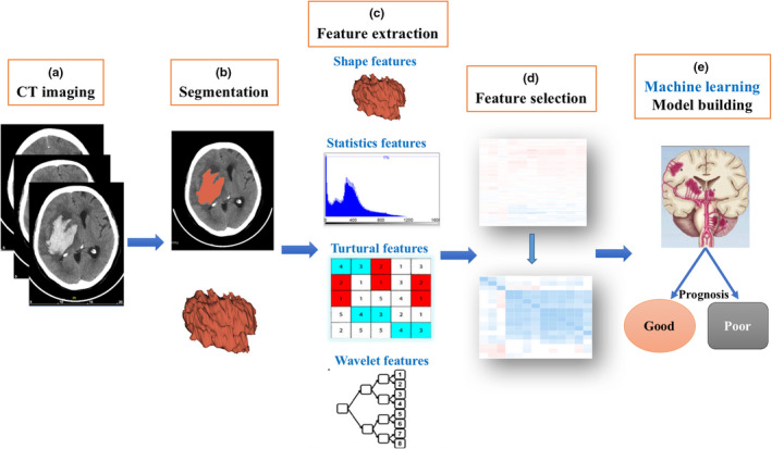
FIGURE 1.

The image postprocessing workflow. First, hematoma on CT images was segmented. After feature extraction, feature selection, and machine learning model construction, six prognosis‐predictive models were established in the training set and were further evaluated in the validation sets
