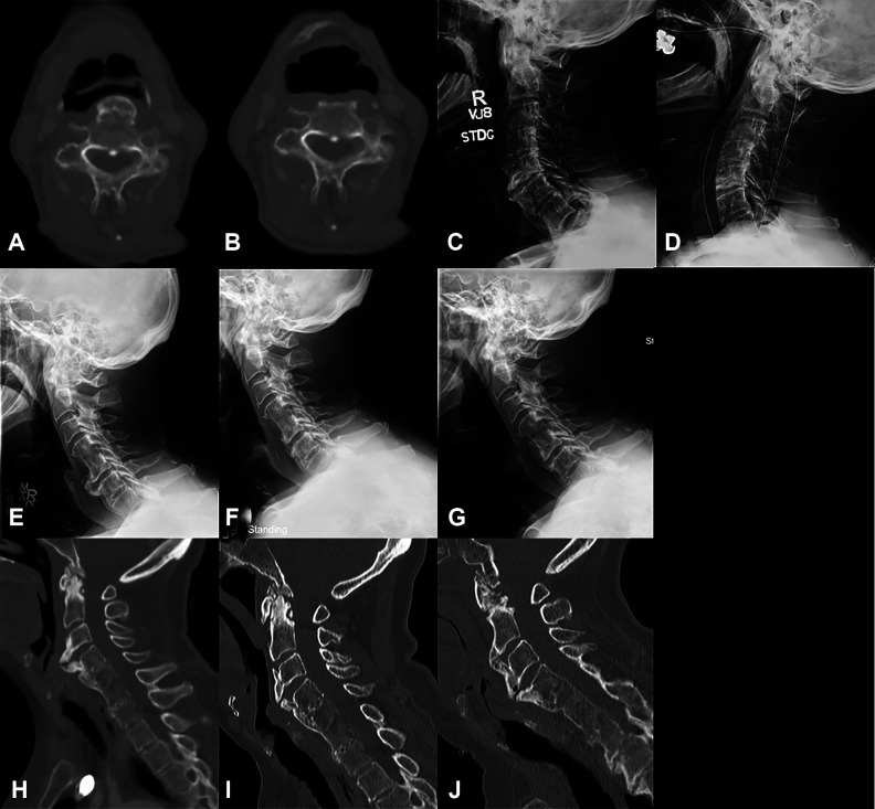
Figure 4.
Imaging of patients who did not see significant improvement in their dysphagia after anterior cervical osteophyte resection. Axial computed tomography (CT) scan through the C4 vertebral body preoperatively (A) and 2 months postoperatively of patient 1 showing minimal residual anterior osteophyte and decreased esophageal compression. Lateral X-rays of patient 13, preoperatively (C) and 1 month postoperatively (D) showing full resection of osteophytes. Radiographic imaging of patient 5 who had a 16-mm osteophyte at C5-6 preoperatively (E), 6 mm postoperatively (F), and osteophyte regrowth to 11.5 mm 2 years postoperatively (G). CT imaging of patient 15 who had a 16.5 mm osteophyte at C3-4 preoperatively (H), 8 mm 14 months postoperatively (I), and regrowth to 13 mm at 6.5 years after osteophyte resection (J).

