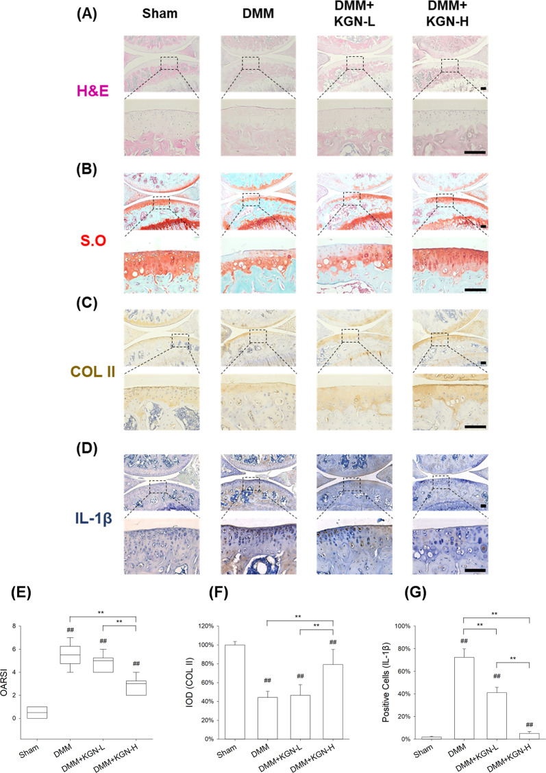
Fig. 3. Effect of intra-articular injection of KGN on the degeneration of cartilage in DMM-induced OA in mice.
DMM-op mice were injected with 1 μM or 100 μM of KGN, twice a week for four weeks, whereas the controls were injected with 10 μL of saline. A–D Representative histological images of medial femoral condyle and tibial plateau of OA mice following immunohistochemical staining. Sagittal sections of cartilage following A hematoxylin and eosin (H&E) staining, B Safranin O (S.O.)/Fast Green staining, C immunohistochemistry for COL II (IHC), and D immunohistochemistry for IL-1β (IHC). (Scale bar = 100 μm). E OARSI scores based on Safranin O/Fast Green staining. F Integrated optimal density (IOD) of COL II in articular cartilage. G Percentage of IL-1β positive cells in articular cartilage. In each section, the quantitative analyses were performed at three random regions, with the average used as the final value. Values represent mean ± S.E.M of six replicas. (*p < 0.05 and **p < 0.01; between the indicated groups and #p < 0.05 or ##p < 0.01 versus the CTRL group).

