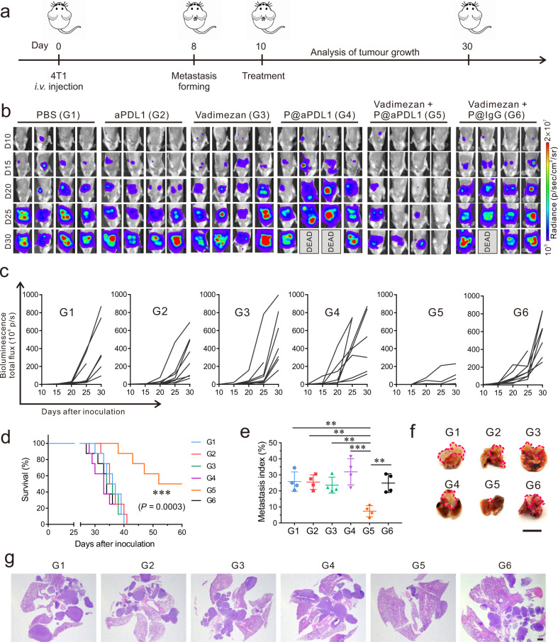
Fig. 3. P@aPDL1 combined with Vadimezan promotes anti-tumour effects in the 4T1 tumour model.
a Schematics of the 4T1 breast cancer metastatic model and treatment. Mice received were treated 10 days post-tumour cells inoculation. Mice received 1 mg kg−1 aPDL1 antibody or 15 mg kg−1 Vadimezan. b In vivo tumour bioluminescence images of mice that received different treatments. c Tumour bioluminescence intensity growth kinetics in different groups. d Survival rates of mice treated in different groups, n = 8 biologically independent animals. e Metastatic index (tumour region/total region) and f representative lung photograph and g H&E staining of tumour lesions after different treatments. n = 4 biologically independent samples in e, pink line indicate the tumours in the lung parenchyma in f. Scale bar for f is 1 cm, scale bar for g is 1 mm. G1, PBS; G2, aPDL1; G3, Vadimezan; G4, P@aPDL1; G5, P@aPDL1 + Vadimezan; G6, P@IgG + Vadimezan. Data are presented as mean ± SD, survival statistical significance was analysed by log-rank (Mantel–Cox) test, and the statistical significance in e was analysed via ANOVA (one-way, Tukey post-hoc test). P-value: **P < 0.01, ***P < 0.001. e G1 vs. G5: P = 0.0011, G2 vs. G5: P = 0.0013, G3 vs. G5: P = 0.0034, G4 vs. G5: P < 0.0001, G6 vs. G5: P = 0.0018.

