Figures 1–4.
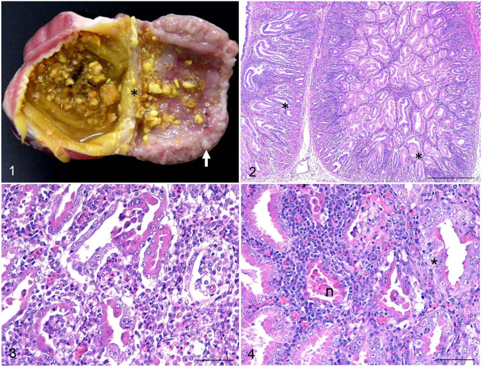
Transmissible proventriculitis in broiler chickens. Figure 1. Longitudinal section of the proventriculus and ventriculus. Dilation of the lumen and isthmus (asterisk) with diffuse thickening of the proventricular mucosa in association with white nodules in the mucosa (arrow). Figure 2. Proventricular mucosa of a 20-d-old broiler with marked inflammatory infiltrate in the interstitium and in the lumen of glands. There is also loss of glandular epithelium and ductal epithelial hyperplasia (*). H&E. Bar = 500 µm. Figure 3. High magnification of Figure 3. Necrosis and loss of glandular cells, replacement of glandular epithelial cells with ductal epithelium (ductal metaplasia), and diffuse infiltrate of lymphocytes and histiocytes in the interstices and in the lumens of acini. H&E. Bar = 50 µm. Figure 4. Proventricular mucosa of a 32-d-old broiler. Marked ductal metaplasia (*) is seen at the base of the gland, associated with epithelial necrosis (n) and infiltration by lymphocytes and histiocytes. H&E. Bar = 50 µm.

