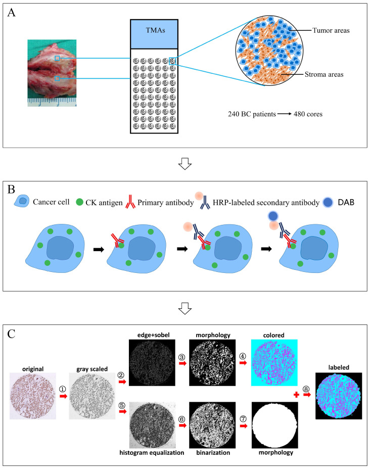
Figure 1.
Major technical procedures of this study. Panel A, TMAs with 480 cores were constructed using 240 cases of breast cancer specimens. Panel B, IHC staining of CK was performed. Panel C, computerized TSR assessment was performed, involving the following steps, ①Transforming the color image into grayscale image; ②Calculating the image gradient using edge and sobel operators to detect the contours of objects; ③Obtaining the tumor objects in the image using morphological operation (dilate-> fill-> erode) with small objects eliminated; ④Staining the tumor objects and non-tumor areas in the image with different colors (colored image); ⑤Increasing the image contrast differences using histogram equalization for subsequent segmentation; ⑥Performing image segmentation using otsu algorithm; ⑦Obtaining the whole core object in the image (mask map); ⑧Merging the colored image and mask map to get the final image and calculating the TSR, TSR=area of stroma objects (in cyan)/area of the whole core object (tumor objects (in magenta) + stromal objects (in cyan)).

