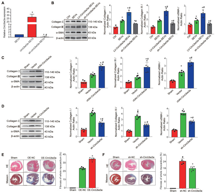
Figure 5.
circUbe3a promotes myocardial fibrosis after AMI. A: Differentially expressed circUbe3a in circUbe3a-SEVs and sh-circUbe3a-SEVs was further assessed by RT-qPCR (*P < 0.05 compared with circUbe3a-SEVs). B: Representative Western blot results from three independent experiments and quantification of collagen I, collagen III, and α-SMA expression in recipient CFs treated with M2M-SEVs in the absence or presence of circUbe3a. (*P < 0.05 versus the Ctrl group; #P < 0.05 versus the SEVs group; n = 3 per group). C: Representative Western blot results from five independent experiments and quantification of collagen I, collagen III, and α-SMA expression in cardiac muscle tissues treated with rAAV-circUbe3a or its NC. (*P < 0.05 versus the sham group; #P < 0.05 versus the vector group; n = 5 per group). D: Representative Western blot results from five independent experiments and quantification of collagen I, collagen III, and α-SMA expression in cardiac muscle tissues treated with sh-circUbe3a or its NC (*P < 0.05 versus the sham group; #P < 0.05 versus the vector group; n = 5 per group). E: Representative micrographs of left ventricular sections (H&E-stained and Masson's trichrome staining) and summary of the semiquantitative analysis of the Masson's trichrome-positive area. (*P < 0.05 versus the sham group; #P < 0.05 versus the OE-NC group; n = 6 per group. Scale bars =200μm). F: Representative micrographs of left ventricular sections (H&E-stained and Masson's trichrome staining) and summary of the semiquantitative analysis of the Masson's trichrome-positive area (*P < 0.05 versus the sham group; #P < 0.05 versus the sh-NC group; n = 6 per group. Scale bars =200μm).

