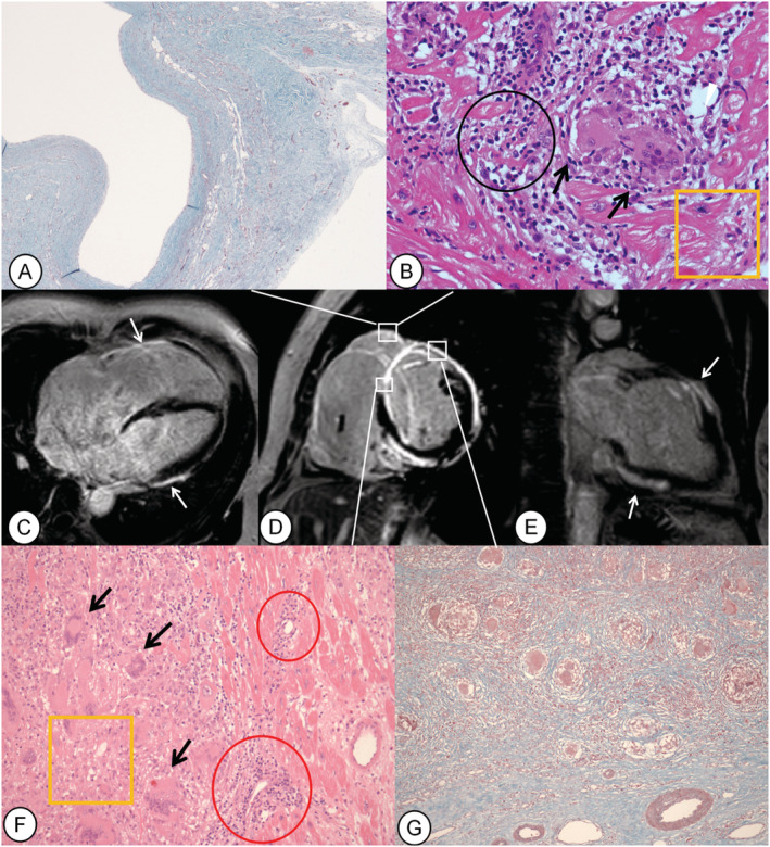
Figure 1.

Female, 51 years of age, presenting chest tightness for more than 1 year and syncope twice in 2 days (Case 4). Late gadolinium enhancement (LGE) images of the short‐axis (D), four‐chamber (C), and two‐chamber (E) views show enhancing area in the right ventricular (RV) wall, transmural and both‐sided LGE in the septum, transmural and epicardial LGE in the anterior wall, and epicardial LGE in the lateral and inferior walls (white arrows). Histopathologic findings show the transmural fibrosis in the RV free wall (A: Masson stain, ×40), anterior septum and anterior wall of left ventricle (G, Masson stain, ×100), and multinucleated giant cells in the anterior wall of left ventricle, septum (F: haematoxylin–eosin stain, ×200), and RV wall (B: haematoxylin–eosin stain, ×400). Black arrows indicate multinucleated giant cells, black circle indicates lymphocytic infiltrate, yellow rectangles indicate damaged myocardium, and red circles represent capillaries surrounded with lymphocytic infiltrates.
