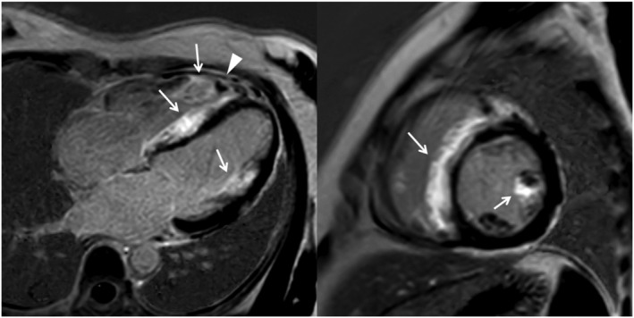
Figure 2.

Four‐chamber view (left panel) and short‐axis view at the mid‐ventricular level (right panel) of late gadolinium enhancement images show the extensive enhancement predominantly involving the right‐sided septum and the anterior papillary muscle (arrows). Besides, there is a right ventricular apical thrombus (arrowheads).
