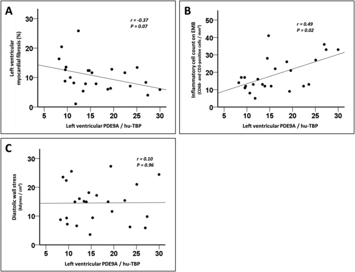
Figure 2.

Association between left ventricular phosphodiesterase 9A (PDE9A) expression, cardiac fibrosis, myocardial inflammation, and diastolic wall stress. (A) In patients with heart failure with preserved ejection fraction (HFpEF), left ventricular PDE9A expression displayed a non‐significant inverse relationship with left ventricular endomyocardial fibrosis, as assessed by histology. (B) Left ventricular myocardial inflammation was analysed by immunohistochemistry in endomyocardial biopsies (EMBs) from patients with HFpEF, and the inflammatory cell count was derived from the total number of CD68‐positive macrophages and CD3‐positive T cells in EMBs. A significant positive correlation between left ventricular PDE9A levels and left ventricular myocardial inflammation was apparent in HFpEF. (C) Left ventricular meridional diastolic wall stress was calculated using haemodynamic and echocardiographic data from patients with HFpEF. No correlation was evident between left ventricular PDE9A levels and diastolic wall stress in patients with HFpEF.
