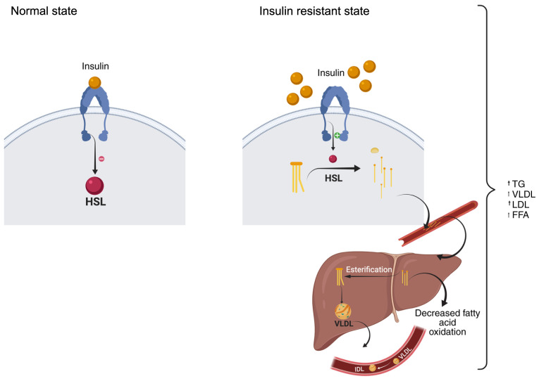
Figure 1.
IR and changes in FA composition. In adipose tissue, in the normal state, insulin binds to the insulin receptor, inhibiting the enzyme HSL in the adipocytes of the adipose tissue. This HSL hydrolyses lipids such as TGs. In the state of IR, insulin is unable to bind to insulin receptors, thereby suppressing intracellular signals. In turn, it activates the HSL enzyme, which hydrolyse TGs to glycerol and FFAs, which are released into the circulation towards the liver. Liver hepatocytes take up FFAs to channel them to their secretory pathways. In the case of hyperinsulinemia/IR, esterification is increased. In the blood vessels, the enzyme LPL hydrolyses monoglycerides and FFAs. A certain proportion of these are delivered to liver LPL (hydrolysis). This process continues and more LDL and FFAs are formed (schematic created with BioRender.com). FFA, free fatty acid; HSL, hormone-sensitive lipase; LPL, lipoprotein lipase; LDL, low-density lipoprotein; VLDL, very low-density lipoprotein; TG, triglyceride; IR, insulin resistance.

