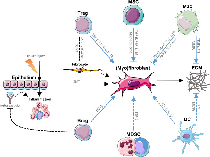
Figure 1.
Direct effects of regulatory immune cells on (myo)fibroblasts and extracellular matrix (ECM) in idiopathic pulmonary fibrosis (IPF). Repeated tissue injury triggering chronic tissue damage resulting in inflammation, epithelial mesenchymal transition (EMT) and ultimately in excessive production and buildup of ECM by fibrocytes and (myo)fibroblasts (fibrosis) in the lungs, represents the main paradigm involved in the pathology of IPF. Immune cells with regulatory functions modulate (myo)fibroblast generation, (myo)fibroblast function and ECM homeostasis through various signaling pathways, and known direct pathways are listed here. Mesenchymal stem/stromal cells (MSCs) promote (myo)fibroblasts through fibroblast growth factor (FGF), transforming growth factor (TGF)-β and interleukin (IL)-1β, while MSC-derived extracellular vesicles (MSC-ECV) and stanniocalcin (SC)-1 have opposite effects. MSCs are also prone to myofibroblastic transition. Tregs promote fibrogenesis through TGF-β, platelet-derived growth factor (PDGF)-B and IL-1β, while inhibiting the recruitment of fibrocytes by inhibition of the CXCL12/CXCR4 axis as well as FGF-9. Macrophages enhance fibrosis through TGF-β, PDGF, tumor necrosis factor (TNF)-α, fibronectin (FN) and inhibit fibroblasts via itaconate and peroxisome proliferator-activated receptor (PPAR) ligands. Myeloid-derived suppressor cells (MDSCs) and regulatory B cells (Bregs) have been suggested to activate lung fibroblasts, possibly through TGF-β. Bregs inhibit autoreactive immunoglobulins, which may deposit in lung tissue and promote inflammation and IPF. Dendritic cells (DCs) have been shown to produce pro-fibrotic TGF-β and IL-1β. Macrophages (Macs) as well as DCs break down the ECM via matrix metalloproteinases (MMPs), a process that is inhibited by tissue inhibitors of metalloproteinases (TIMPs) produced by other macrophage subtypes. Both Macs and DCs have been found to produce fibronectin (FN), another ECM component. Blue lines represent the direct effects of regulatory immune cells on (myo)fibroblasts and ECM in IPF. Dashed lines represent interactions that are not firmly established in IPF. This figure was created using Servier Medical Art templates, which are licensed under a Creative Commons Attribution 3.0 Unported License; https://smart.servier.com.

