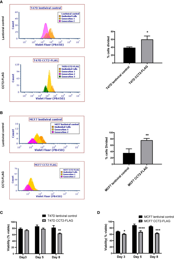
Figure 7.
CCT2 overexpression increases cell division of breast cancer cells in 3D cultures. (A, B) The ViaFluor® dye was used to assess cell division over time for T47D cells (A) and MCF7 cells (B), CCT2-FLAG overexpressing and lentiviral controls, from days 3-5 of spheroid 3D cultures are shown. Cells were stained at day 3 and incubated for 48 hours before assessing cell division by flow cytometry. Generation time (histograms) and percent cells divided (graph) was determined (n=3). (C, D) PI exclusion assay was used to assess the viability of cells from spheroid cultures at days 3, 5, and 8 of 3D growth for T47D cells (C) and MCF7 cells (D) (n=3). Data was acquired using a CytoFlex S flow cytometer and analyzed using FCS Express software. Values are mean with SD. *p-value <0.05, **p- value <0.005, ***p-value <0.0005, ****p-value < 0.00005.

