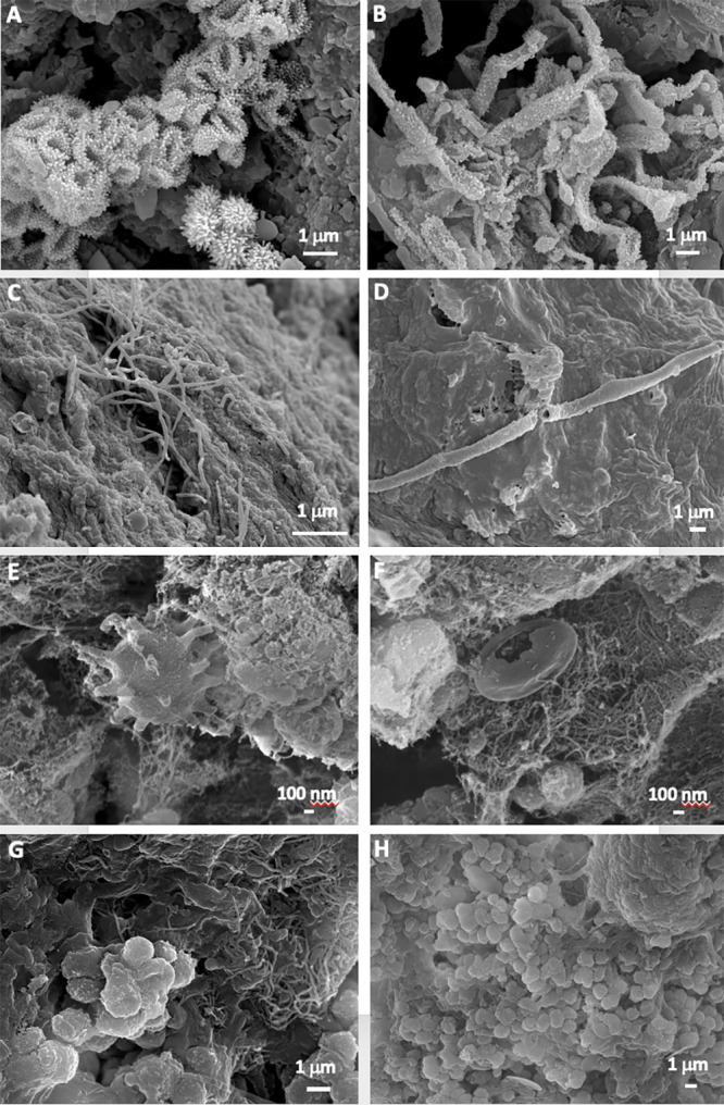
Figure 3. Field Emission Scanning Electron Microscopy images of samples.

Representative Field Emission Scanning Electron Microscopy images of the studied samples, depicting (A,B) Clusters of Actinobacteria-like cells with spiny surface ornamentation in the mucous formation of ochre color (MZ03-2B); (C) Microbial filaments probably of Actinobacteria in the ochre soft stalactite (MZ03-3C); (D) Hollow bacterial filament on the sample surface of the ochre soft stalactite (MZ03-3C); (E) Prosthecate-like bacterial cell in the ochre mucous deposit on a rock crack with water runoff (MZ03-8H); (F) Dense network of nano-scaled filaments in the ochre mucous deposit on a rock crack with water runoff (MZ03-8H); (G) Mass of interwoven filaments, and (H) Coccoid-shaped cells with smooth surfaces in the mineral formation with abundant moonmilk deposits (MZ03-10J).
