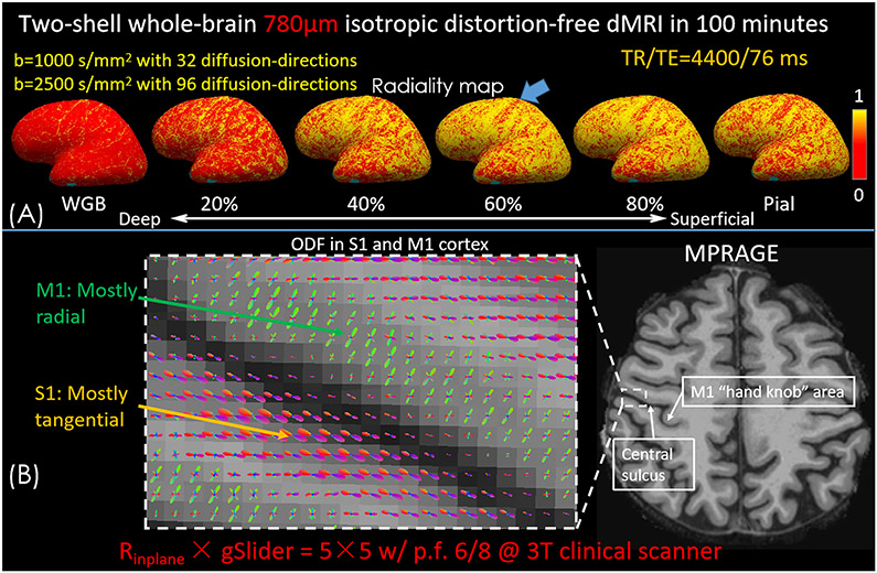
Figure 6.
(A) Radiality maps at the surface of the white-gray boundary (WGB) surface, the surfaces of 20%, 40%, 60% and 80% of cortical thickness into gray matter, and the pial surface on the inflated brain surface. (B) ODF results in primary motor cortex (M1) and somatosensory cortex (S1) obtained from the whole brain 780μm dMRI data with two shells.

