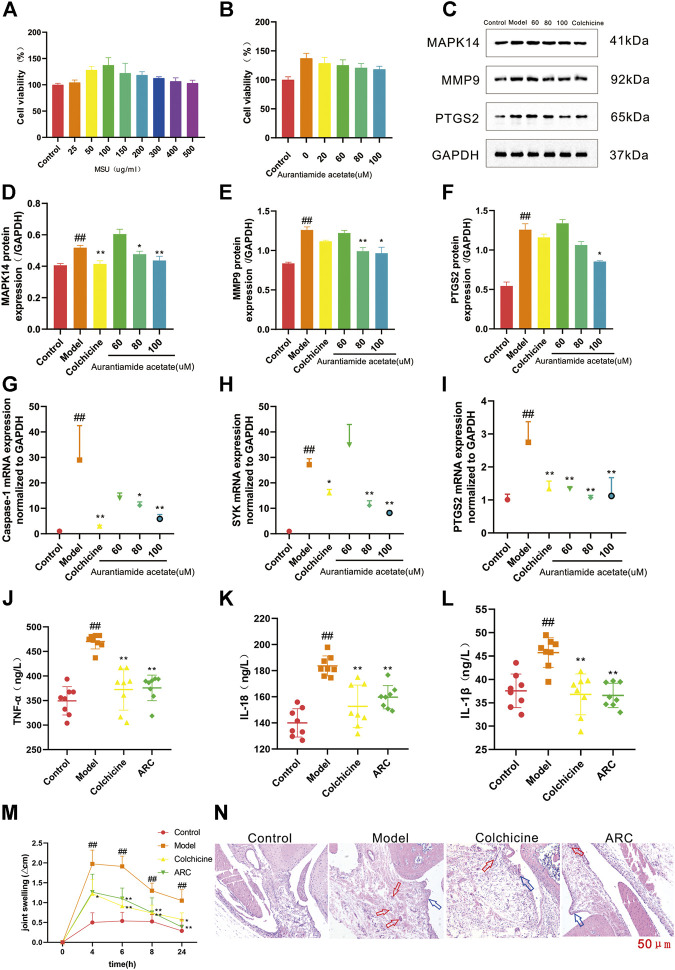
FIGURE 6.
(A) THP-1 cells were exposed to MSU at various concentrations for 24 h. (B) Protective effects of AA on the viability of MSU-induced THP-1 cells. (C) Western blotting. (D) Expression of MAPK14 protein. (E) Expression of MMP9 protein. (F) Expression of PTGS2 protein. (G) Expression of caspase-1 mRNA. (H) Expression of SYK mRNA. (I) Expression of PTGS2 mRNA. (J-L) Effect of ARC on inflammatory factors in rat serum. (J) TNF-α. (K) IL-18. (L) IL-1β. (M) The knee joint circumference of rats were determined at 0, 4, 6, 8, 24 h after MSU stimulation, the increment at different time point was calculated. (N) Histopathological analysis of rat synovium tissue (100×). The red arrows pointed to inflammatory cells, blue arrows pointed to synovial hyperplasia. #p < 0.05, ##p < 0.01 vs. control group; *p < 0.05, **p < 0.01 vs. model group.

