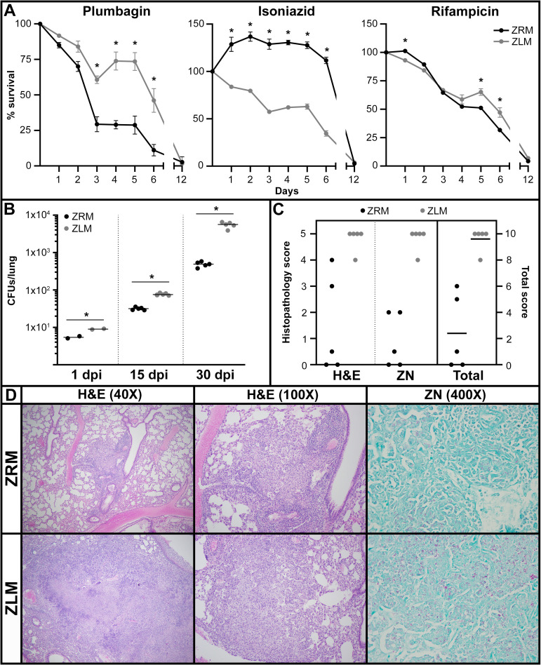
Fig 5. Zn2+-limited Mtb exhibit increased resistance to oxidative stress in vitro and cause higher bacterial burden and pathology in vivo.
(A) Survival of Mtb mc2 6206 after growth in ZRM or ZLM and subsequent exposure to the indicated chemical or antibiotic. Survival was monitored with flow cytometry for twelve days following treatment and was calculated as the percentage of live cells in untreated cultures at the beginning of treatment (S10 Fig). The data are representative of three independent experiments and are given as the average of biological replicates (ZRM n = 3, ZLM n = 2) with error bars representing standard deviation. Asterisks represent a statistically significant difference (t-test, p-value <0.05) between survival of cultures in ZRM vs. ZLM at any given time-point. (B) Bacterial burden in the lungs of C3HeB/FeJ (Kramnik) mice after 1, 15 and 30 days post-infection (dpi) with Mtb H37Rv pre-grown in ZRM or ZLM. Horizontal bars across the data points represent the average CFUs. Asterisks represent a statistically significant difference (t-test, p-value <0.05) between CFUs of mice infected with ZRM vs. ZLM at each timepoint. (C) Blinded histopathology scores from hematoxylin and eosin (H&E) and Ziehl-Neelsen (ZN) staining in panel D. Maximal score of five for H&E and ZN stained cross sections is based on observed changes in lung morphology and immune cell infiltration and bacterial burden respectively and these scores are plotted on the left y-axis. The total score is the sum of the H&E and ZN score for each lung and is given on the right y-axis with a score of 10 representing the maximal level of disease pathology. The horizontal bar through the data for total score represents the average total pathology score. The non-parametric Mann-Whitney U-test for significance was applied to histopathology scores from H&E, ZN and total, all resulting p-values <0.02 indicating a significant difference in pathology scores for mice infected with Mtb pre-grown in ZRM or ZLM. (D) Histopathology micrographs of the lungs of mice infected with Mtb H37Rv pre-grown in ZRM or ZLM at 30 days post-infection. In blinded studies, histology cross-sections of lung tissues were stained with H&E to score changes in lung morphology and immune cell infiltration or ZN stain for mycobacteria (pink) to assess bacterial burden.

