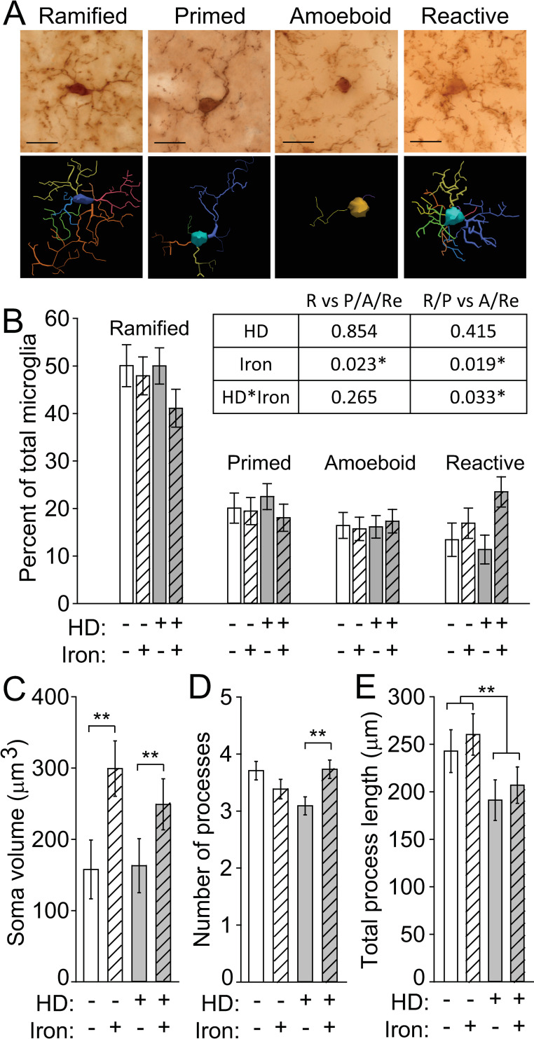
Fig 4. Neonatal iron supplementation and HD differentially alter microglial morphology.
Microglia in brain sections from 14-week-old N171-82Q mice were labeled with anti-Iba1, and their morphology was evaluated. A. Representative micrographs of microglial cells labeled with anti-Iba1 were identified and characterized as having one of four morphologies, amoeboid, primed, ramified, and reactive. Neurolucida traces are shown below each image. Scale bars = 20 μm. B. Iron increased the frequency of reactive microglia in iron-supplemented HD mice. The table shows p-values from logistic regression analysis of microglial morphologies. Iron significantly increased the probability of activated microglial morphologies. R = ramified; P = primed; A = amoeboid; Re = reactive. Each morphology is presented as a percent of total microglial cells within each mouse. The percentage of cells with each morphology relative to all microglial cells in a given mouse are shown. C. The microglial soma volume was significantly increased by NIS. D. Iron-supplemented HD mice have a significantly increased number of processes relative to vehicle-treated HD mice. E. The total process length was significantly decreased in HD mice relative to WT mice but was not changed by iron supplementation. B-F. n = 10 WT control, 11 WT NIS, 12 HD control, 11 HD NIS. Data are shown as the mean ± SE. *p<0.05, **p<0.01.

