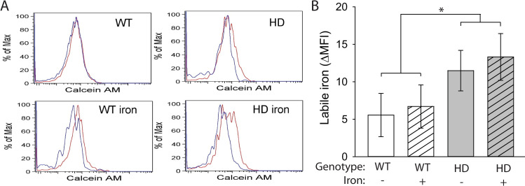
Fig 5. Labile iron accumulates in HD microglia.
Microglia, defined as CD11b+CD45+CX3CR1+ cells, were extracted from mouse brains at 14 weeks of age and labeled with calcein AM. A. Representative histograms of calcein AM fluorescence in brain microglia incubated with (red) or without (blue) deferiprone. B. Labile iron, defined as the difference in mean fluorescence intensity (MFI) between cells incubated with and without deferiprone, was significantly increased in HD microglia. n = 6–8. Data are shown as the mean ± SE. *p<0.05.

