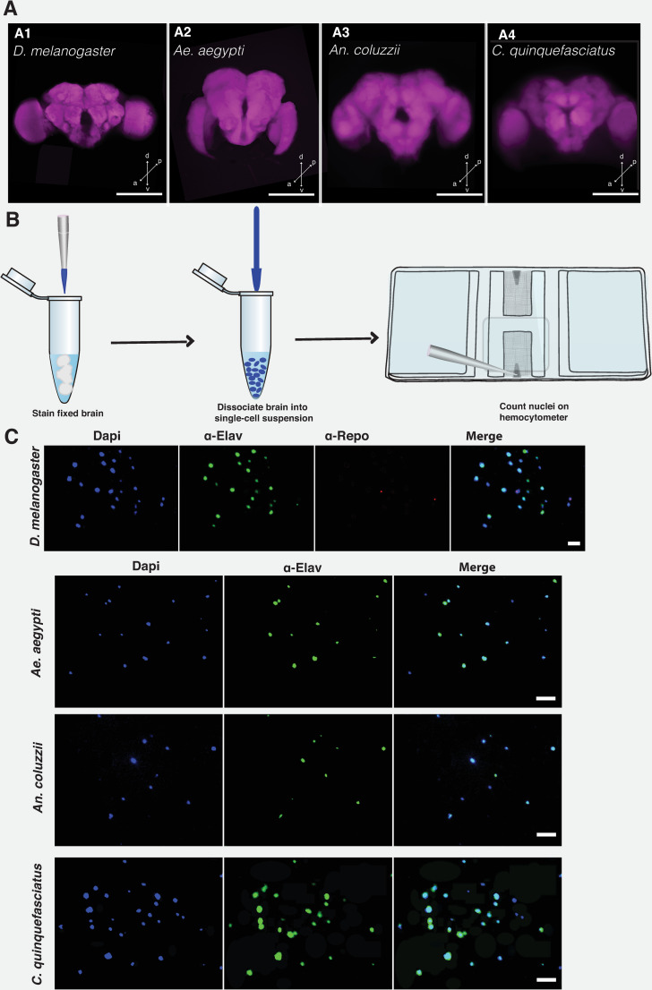
Fig 1. Estimating brain cell population using isotropic fractionator method.
Confocal Z-stacks of the whole brain of D. melanogaster (A1), Ae. aegypti (A2), An. coluzzii (A3), and C. quinquefasciatus (A4). Brain was counterstained with nc82 antibody that labels the pre-synaptic active zone protein, Bruchpilot. Whole brain was imaged on a LSM 700 Zeiss confocal microscope at 512 × 512 pixel resolution, with 1.04 μm pixel size. Scale bar represents 50 μm. Arrow directions represent the dorsal (d), ventral (v), posterior (p), and anterior (a), respectively. (B). Cartoon showing the steps for performing isotropic fractionator. Brain tissue is fixed and stained with the appropriate dye or antibody. Tissue is dissociated into a single-cell suspension, which is then counted on a Neubauer counting chamber or hemocytometer. (C) Images of nuclei are stained with DAPI (4’, 6-diamidino-2-phenylindole dihydrochloride), α-elav (embryonic lethal abnormal vision), and α-repo (Reversed polarity protein) to label nuclei, neuronal and non-neuronal cells, respectively. Staining of glial cells with α-repo was possible only for Drosophila brain cells. Scale bar represents 10 μm. For illustration purposes, all images were processed on ImageJ software and Adobe Illustrator.

