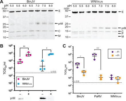
Fig. 3. Maturation of BinJV and WNVKUN particles by in vitro furin cleavage.

(A) Purified BinJV (left) and immature WNVKUN (right) were incubated with furin in buffer ranging from pH 5 to 8 and analyzed by nonreducing SDS-PAGE and visualized by SYPRO Ruby stain. Viral proteins indicated by arrows. (B) Infectivity of BinJV and immature WNVKUN virions in C6/36 cells with or without furin treatment. Cleavage was analyzed by SDS-PAGE, and virus titers were determined by TCID50. n = 3 biological replicates, and statistical analysis was performed by two-stage multiple t tests against nontreated (furin negative). *P ≤ 0.05 and **P ≤ 0.01. Means ± SD. Limit of detection (L.O.D.) for TCID50 is 2.3 log10 TCID50 ml−1. (C) Infectivity of BinJV, PaRV, and WNVKUN in C6/36 cells pretreated with or without 25 μM furin inhibitor (FI). Virus titers were determined by TCID50; n = 3 biological replicates, and statistical analysis was performed by two-stage multiple t tests against nontreated (FI negative). *P ≤ 0.05 and ***P ≤ 0.001. Means ± SD. Limit of detection for TCID50 is 2.3 log10 TCID50 ml−1.
