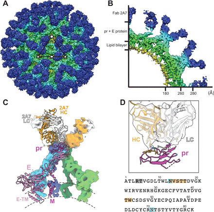
Fig. 6. Cryo-EM reconstruction of BinJV:2A7.

(A) Surface and (B) wedge cross-section of the cryo-EM reconstruction of BinJV:2A7-Fab. Map was colored according to radius (red, 0 to 30 Å; yellow, 31 to 140 Å; green, 141 to 180 Å; cyan, 181 to 280 Å; and blue, >280 Å). (C) Fit of a homology model of the 2A7-Fab and the structure of BinJV within the reconstruction of the complex. Dimers of prM-E are colored according to symmetry position: twofold with green/lime for prM/E, threefold with blue/cyan for prM/E, and fivefold with magenta/pink for prM/E. The HC and LC of the 2A7-Fab is represented in orange and white, respectively. Surfaces for one member of the asymmetric unit are omitted to show a cartoon representation. (D) Zoom of the pr:2A7 interface with the sequence of the N-terminal domain of pr displayed below. Residues of pr that are within 7 Å of the Fab are indicated by dark orange (HC) and gray (LC). N-linked glycan sites of pr are highlighted in light blue.
