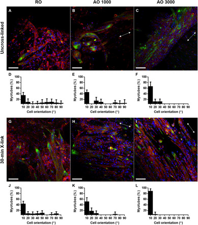
Fig. 5. Alignment of myotubes formed on dECM scaffolds.

Immunofluorescence staining of myotube formation [desmin (green), actin (red), and nuclei (blue)] was captured via confocal microscopy. (A to C and G to I) Representative images of myotube formation and (D to F and J to L) the orientation graphs of myotubes grown on cell-laden dECM constructs after 7 days in induction media are shown. Scale bar represents 100 μm and is the same across images. The white double-headed arrow represents the direction of myotube alignment. Values represent means ± SD (n = 9).
