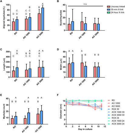
Fig. 6. Analysis of myotube formation on dECM scaffolds.

Myotube formation was assessed after 7 days of culture in induction media. (A) Myotubes aligned within 10° of the origin expressed as a percentage of all myotubes, (B) fusion index (nuclei per myotube), (C) myotube length, and (D) myotube width were assessed from confocal images. (E) Myotubes per image were counted and reported for each group. (F) In vitro scaffold degradation was assessed throughout the 11 days that cell-laden scaffolds were cultured. Samples were cultured in growth media during days 0 to 4 and then switched to induction media during days 4 to 11. Values represent means ± SD [n = 9 (A to E) and n = 3 (F)]. Bars that share letters are not significantly different. Conversely, bars that do not share a letter are significantly different (P < 0.05). n.s. indicates no significant difference between all groups (P < 0.05).
