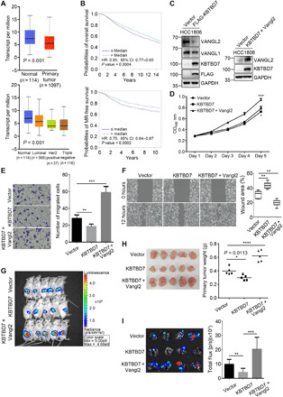
Fig. 7. KBTBD7 inhibits growth and metastasis of mammary tumors.

(A) TCGA data analysis of breast cancer. KBTBD7 was down-regulated in breast carcinoma compared to normal breast tissues, Student’s t test, P < 0.001. (B) Kaplan-Meier survival analysis of 4295 patients for overall survival and 2637 patients for metastatic relapse–free (MR-free) survival (univariate Cox analysis, P = 0.0004 and 0.0002). Patients with lower KBTBD7 expression level have decreased overall survival and MR-free survival. HR, hazard ratio. CI, confidence intervals. (C) Establishment of a HCC1806 breast cancer cell line stably expressing FLAG-tagged KBTBD7 (HCC1806-KBTBD7, left). Vangl2 was further stably expressed in HCC1806-KBTBD7 cells (right). (D) XTT assays of HCC1806 stable cancer cell lines. Expression of KBTBD7 slightly inhibited but further expression of Vangl2 promoted the proliferation of HCC1806 cells. (n = 3 repetitions; Student’s t test, *P < 0.05, ***P < 0.001). (E and F) Transwell migration (E) and wound healing (F) assays of HCC1806 stable cancer cell lines. KBTBD7 inhibited but Vangl2 promoted the migration of HCC1806 cancer cells (n = 3 repetitions; Student’s t test, **P < 0.01, ***P < 0.001). (G and H) KBTBD7 expression slightly inhibited but Vangl2 expression strongly promoted tumor growth in mouse xenograft models. (G) Ex vivo luciferase–based bioluminescence imaging of NOD SCID female mice 25 days after injection of HCC1806 stable cancer cells into the fat pads (n = 5 for each group). (H) Primary mammary tumors isolated from NOD SCID female mice 25 days after injection (left). Their body weights were measured (right) (n = 5 for each group; Student’s t test, *P = 0.0113, ****P < 0.0001). (I) Luciferase-based bioluminescence imaging (left) and quantification (right) of the lungs of NOD SCID female mice 8 weeks after injection of HCC1806 stable cancer cells into mammary fat pads (n = 4 for each group, Student’s t test, **P < 0.01, ***P < 0.001). KBTBD7 expression suppressed but Vangl2 expression strongly promoted lung metastasis of HCC1806 breast cancer cells.
