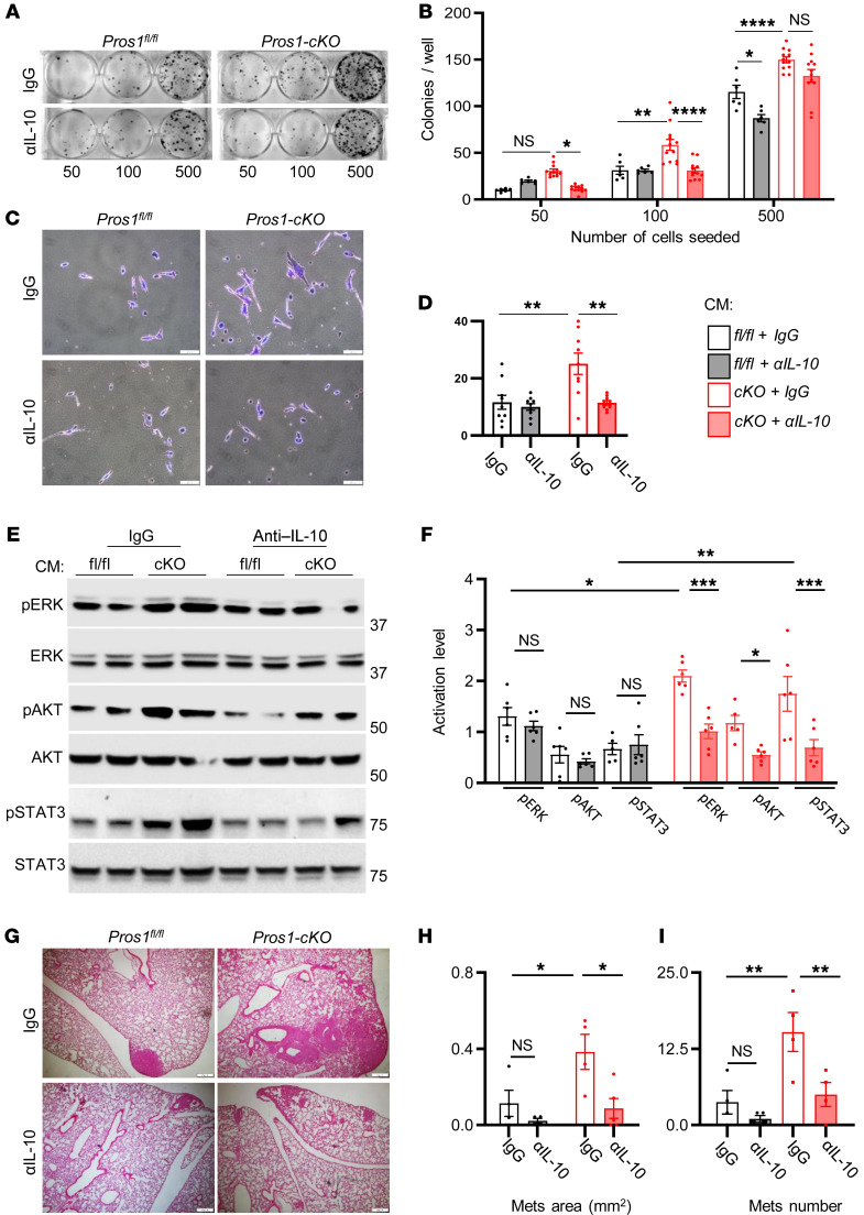
Figure 8. Neutralization of IL-10 in Pros1-cKO conditioned medium reduces Lewis lung carcinoma aggressiveness and suppresses in vivo metastasis.
Lewis lung carcinoma (LLC) cells were educated by the indicated CM with control (IgG) or anti–IL-10 neutralizing antibodies (αIL-10) for 24 hours and subject to a battery of in vitro assays. (A and B) Colony survival after 10 days. Representative images of educated cells plated at different densities as indicated (A) and quantification of colonies (B). Mean ± SEM; n = 6–12 mice/group; 3 independent experiments. *P = 0.02; **P = 0.004; ****P < 0.0001; 2-way ANOVA. (C and D) Matrigel invasion assay. Representative images (C) and quantification (D) of educated cells that traversed the ECM-coated membrane without or with IL-10 neutralization. Scale bars: 50 μm. (D) The average cell number from 7–9 different fields ± SEM is shown, 4–5 mice/group, 2 independent experiments. **P = 0.0017 (left) and 0.0014 (right); 2-way ANOVA. (E) Representative Western blots and quantifications (F) of ERK, AKT, and STAT3 activation from educated LLC cell lysates. Band intensities were calculated from 5–6 mice/group, 2 independent experiments. The average ratio (phosphorylated/total) ± SEM is plotted. *P = 0.04 (ERK), **P = 0.002 (STAT3) for control versus cKO with IgG; and *P = 0.05 (AKT), ***P = 0.001 (ERK and STAT3) for cKO with IgG versus cKO with αIL-10; 2-way ANOVA. (G–I) Educated LLC cells described above were s.c. injected into the flank of WT mice and 3 weeks later lung metastasis was evaluated. (G) Representative images of H&E-stained sections from lungs. Scale bars: 200 μm. (H and I) Scoring of lung metastases (Mets), the average metastatic area (H) and number (I) ± SEM are shown. *P = 0.04 and 0.02 for H (left and right, respectively); **P = 0.01 and 0.008 for I (left and right, respectively); 2-way ANOVA. NS, nonsignificant.

