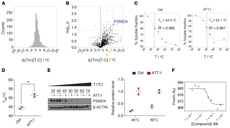
Figure 3. PSMD4 is identified as a molecular target of ATT-I in the immunoproteasome.
(A–D) Cellular thermal shift assay was conducted to identify potential molecular targets of ATT-I in MC38 cells using melting temperature (Tm) shifts. (A) Distribution plots of Δ(Tm) values for proteins from control and ATT-I–treated cells. (B) Volcano plots of Δ(Tm) values to identify potential targets of ATT-I with the most significant melting temperature changes. PSMD4 is indicated on the plots. (C) Temperature based protein-nondenaturation curves for PSMD4 in control and ATT-I–treated cell lysates. (D) Quantitative data from (C) are presented as mean ± SD of 2 parallel experiments (n = 2). Unpaired 2-tailed t test was used for statistical analysis. (E) Representative immunoblots of PSMD4 in the MC38 cell lysates with or without ATT-I treatment are shown. (F) Microscale thermophoresis (MST) binding assay determined the Kd value (Kd = 0.4 μM) for the binding of ATT-I toward PSMD4. Data shown are representative of 4 independent experiments. *P < 0.05.

