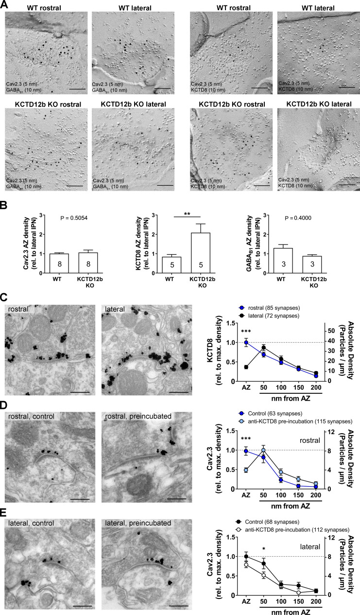
Figure 7. Absence of KCTD12b leads to a compensatory increase of KCTD8 inside the active zone of ventral MHb terminals.
(A) Example images of active zones containing Cav2.3 and either GABAB1 (left) or KCTD8 (right) in replicas of WT (upper row) and KCTD12b KO IPN tissue (lower row). Scale bars: 100 nm. (B) Quantification of relative densities for Cav2.3, KCTD8, and GABAB1 in active zones located in the rostral IPN of WT and KCTD12b KO mice. Densities were normalized to the average density in MHb terminals inside the lateral IPN of the same replica. The number inside the bars indicates the number of replicas used for quantification. **p<0.01 in a Mann–Whitney test. (C) Pre-embedding EM labeling for KCTD8 in KCTD12b KO mice (data from three mice). The density of KCTD8-labeled gold particles was significantly higher in the AZ of ventral MHb terminals in the rostral IPN compared with dorsal MHb terminals in the lateral IPN. ***p<0.0001 two-way ANOVA with Bonferroni post hoc test. (D, E) Comparison of pre-embedding EM labeling for Cav2.3 in MHb terminals in KCTD12b KO mice of control sections (standard labeling, four mice) with sections pre-incubated with anti-KCTD8 primary antibody and biotinylated secondary antibody (three mice). Pre-incubation resulted in significant reduction in labeling densities for Cav2.3 selectively in the active zone of ventral MHb terminals in the rostral IPN as well as in the peri-synaptic region of dorsal MHb terminals in the lateral IPN. ***p<0.001, *p<0.05 two-way ANOVA with Bonferroni post hoc test.

