Fig. 1.
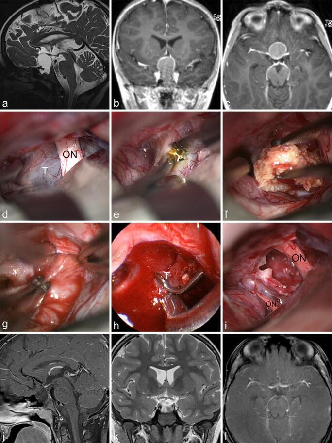
The 7-year-old boy presented with signs of hypopituitarism such as loss of weight, arrest of growth, impairment of his general condition, and fatigue. Since 4 weeks he had been complaining about headache and vomiting. The endocrinological evaluation demonstrated panhypopituitarism, but no obvious DI. a–c MR imaging revealed an intra-suprasellar circumferentially contrast enhancing cystic lesion which was highly suspicious of a craniopharyngioma. d The tumor (T) was approached in the interoptic window via a right-sided frontolateral approach exposing the right optic nerve (ON). e The tumor capsule was incised with a knife. f Calcified tumor parts were removed with tumor forceps. g The tumor was dissected using the bimanual traction-countertraction technique with two forceps. h Intrasellar tumor parts (T) which could not be visualized with the microscope were removed under endoscopic view of a 30° endoscope. i The final inspection showed the gross total tumor resection and intact optic nerves (ON). j–l MR imaging obtained 2 years after surgery showed no recurrence. The boy is doing well under full hormonal replacement therapy

