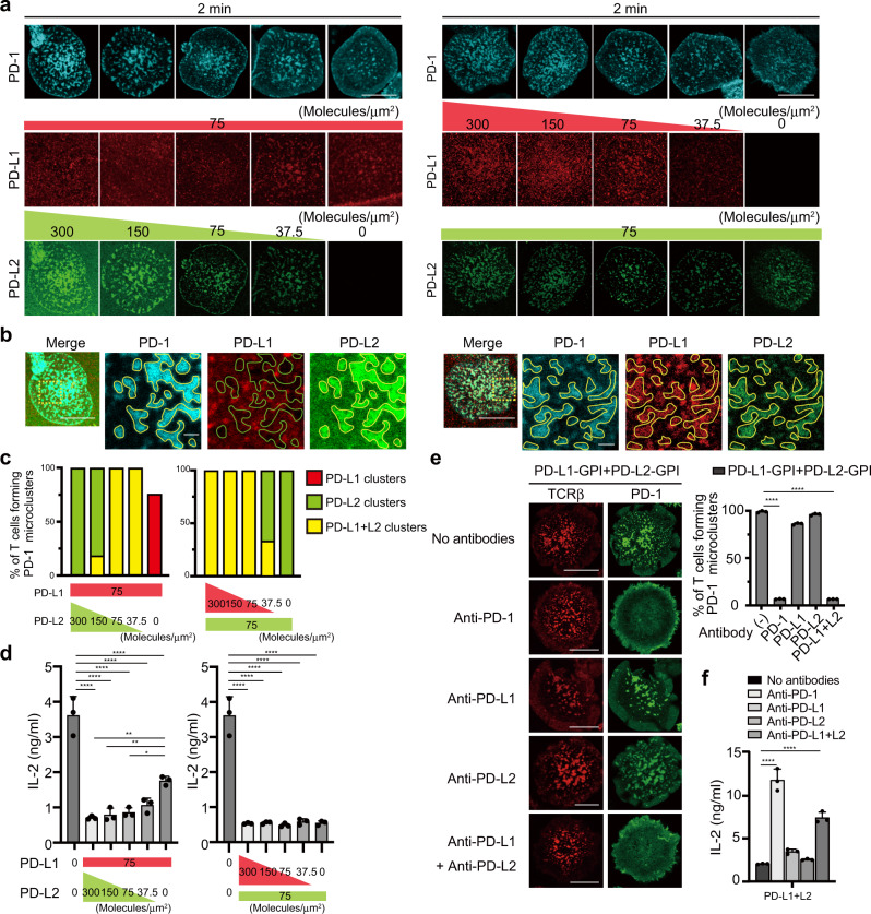
Fig. 5. PD-L2 outcompetes PD-L1 in binding to PD-1 at the TCR–PD-1 microclusters.
a The mPD-L1–GPI (red, middle) and mPD-L2–GPI (green, bottom) were labeled with AlexaFluor 647 and AlexaFluor 488, respectively, and reconstituted into SLBs as in Fig. 1a in the different densities indicated above the images. 2D12 expressing mPD-1–HaloTag (top, cyan) were preincubated with HaloTag ligand–TMR and real-time imaged by confocal microscopy at 2 min after the T cell-bilayer contact. b Images of cells in a at mPD-L1–GPI (75 molecules/μm2) + mPD-L2–GPI (300) (left) or mPD-L1–GPI (300) + mPD-L2–GPI (75) (right). The yellow squares in the left panels are magnified in the right three panels. Yellow enclosures show PD-L1 or PD-L2 clusters colocalized with PD-1 microclusters. Green enclosures show blank where PD-1 microclusters formed. c The graphs show the percentage of T cells forming PD-1 microclusters composed of either PD-L1, PD-L2, or PD-L1 + L2 at the different densities of mPD-L1–GPI and mPD-L2–GPI in a. d Silica beads were coated with the same SLBs as in a with different densities of mPD-L1–GPI and mPD-L2–GPI and prepulsed with MCC88–103 to use as engineered antigen presenting cells. 2D12 expressing mPD-1 were stimulated by these silica beads for 16 h and the concentration of IL-2 in each supernatant was measured by ELISA. e 2D12 expressing mPD-1–EGFP (green) were prestained with DyLight 650-labeled H57 Fab (red) and real-time imaged on an SLB with both mPD-L1–GPI and mPD-L2–GPI in the absence or presence of each antibody as in Fig. 2a. The graph shows the percentage of T cells forming PD-1 microclusters (n = 30). f The cells in e were stimulated by 16h-aggregation culture with 10 μM of MCC88–103 and the concentration of IL-2 in each supernatant was measured by ELISA. All data are representatives of three independent experiments. Bars, 5 μm. Error bars, SD. Statistical analysis was by one-way analysis of variance (ANOVA). *p < 0.05, **p < 0.01, ****p < 0.0001.

