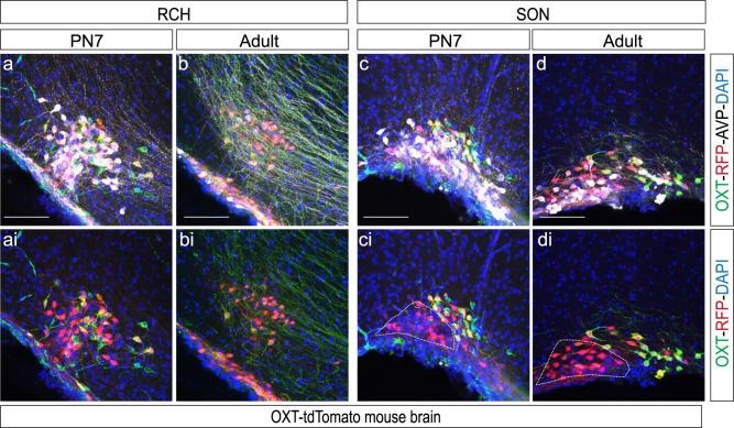
Fig. 9. Development of SON and RCH in an OXT-tdTomato mouse line.
Brain coronal sections show the RCH (a, ai, b, bi) and SON (c, ci, d, di) at a range of stages: PN7 (a, ai, c, ci) including adult (b, bi, d, di). Immunohistofluorescence against anti-OXT (green), anti-RFP (red), and anti-AVP (white). White line represents the population of RFP positive cells not recognized by the PS38 OXT antibody. Abbreviations: SON supraoptic nucleus, RCH retrochiasmaticarea. Scale bar: 100 µm.

