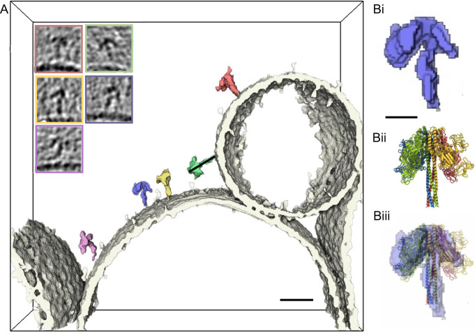
Fig. 3. Vip3act directly visualised on liposome membranes.
Segmented density from cryo-ET shows interaction of Vip3Bc1act (coloured) with the LUV membrane (white). Images inset show section through the tomogram of the matching particle in segmentation. Full tomogram can be visualised in Supplementary Movie 1. Scale bar 20 nm. B Scaled comparison between segmented density for a single particle in the tomogram, with 5 nm scale bar (i) compared to Vip3Bc1act model (ii) with an overlay showing the stalk density fits the 4-helix bundle well, and the density which is distal to the membrane is consistent with single particle Vip3Bc1act model. The helical stalk modelled in Vip3Bc1act does not account for all of the stalk density observed in the sub-volume.

