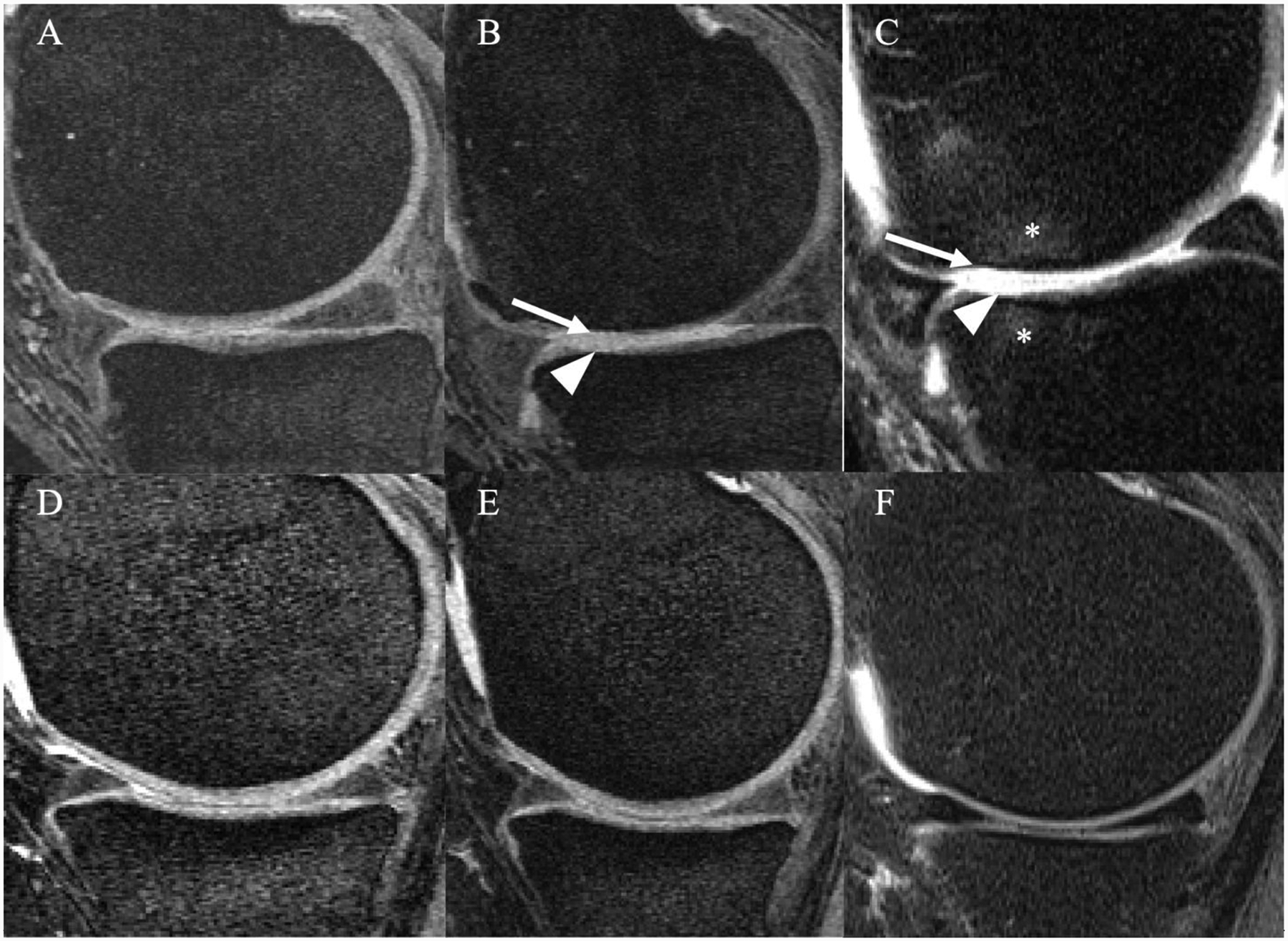
Figure 4:

MR images of the right knee obtained with a sagittal dual-echo steady state (DESS) (A,B,D,E) and intermediate-weighted fat suppressed sequence (C,F) at baseline (A, D) and after 48 months (B, C, E, F). Overweight 64-year-old man in the racquet sports group (A-C) and overweight 47-year-old woman in the elliptical trainer group (D-F). The man in the racquet sports group developed full thickness cartilage defects in both medial femur condyle and tibia in B-C (arrow and arrowhead) with associated bone marrow edema like lesions (asterisks). In contrast, no cartilage defects were seen in the woman in the elliptical trainer group (D, E or F).
