Fig. 2.
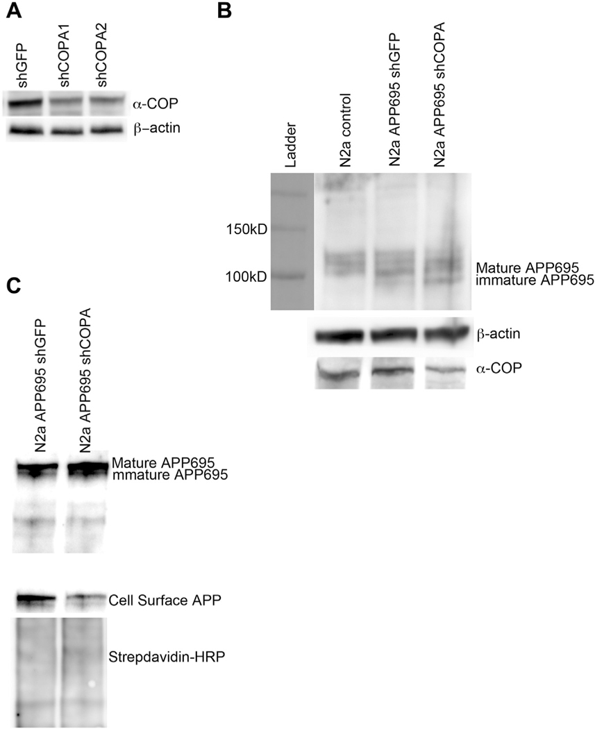
Depletion of α-COP alters processing of APP in N2a cells. (A) Representative Western blot from whole cell N2a lysates after puromycin selection and clonal isolation. Blotting with H3 antibody shows decreased levels of α-COP protein compared with β-actin loading controls. (B) Representative Western blot of whole cell lysate blotted with antibody 22C11 to visualize mature and immature forms of APP695 shows increased levels of immature APP695 in cells with stable knockdown of COPA. (c) Blotting of whole cell lysates and streptavidin-isolated proteins cell surface protein with human-specific APP antibody (E8B3O) shows decreased APP695 at the plasma membrane in COPA knockdown cells compared with controls. Blotting with HRP-conjugated Streptavidin confirms even loading of the cell surface proteins.

