Fig. 3.
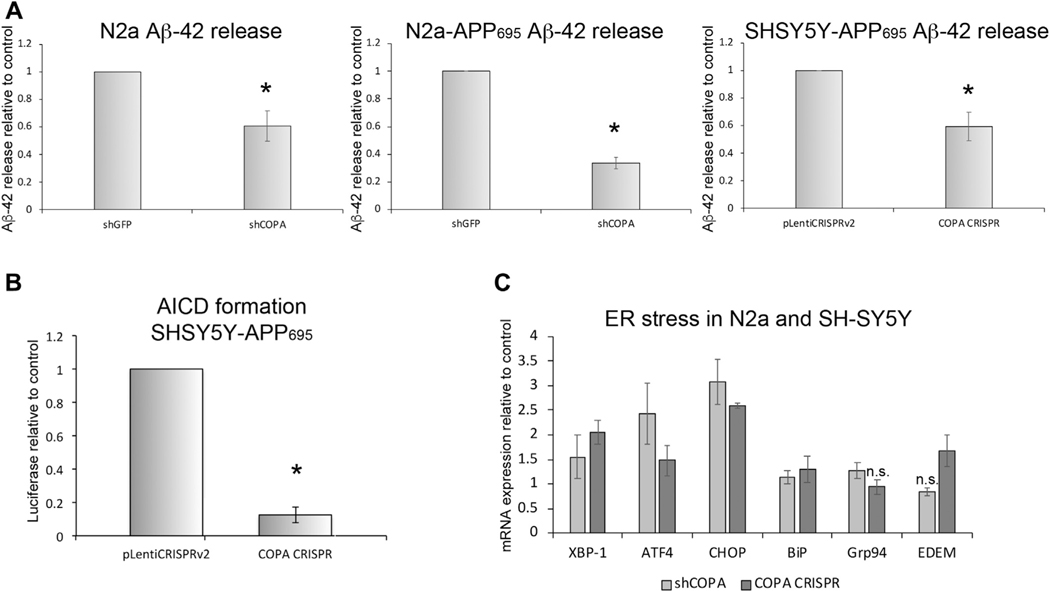
Depletion of α-COP reduces the release of APP fragments. (A) Combined results of ELISA measurements of murine Aβ−42 in N2a cells show decreased Aβ−42 in cell culture media from a clonal line with stable expression of shRNA against COPA (shCOPA) compared with a control clone expressing shRNA against GFP (shGFP). Similar results were detected from a clonal line of N2a cells expressing stably expressing human APP695 in addition to the shRNA. Combined results of ELISA measurements of Aβ−42 in SH-SY5Y cells show a similar significant decrease in Aβ−42 from cells where the level of α-COP protein has been reduced by CRISPR editing of the COPA gene. (B) This graph depicts the combined results of multiple luciferase assays in SH-SY5Y cells expressing a reporter plasmid to measure the formation of the AICD following cleavage of APP695 by γ-secretase. Reducing α-COP protein by CRISPR gene editing resulted in decreased luciferase signal compared with control cells expressing the pLentiCRISPRv2 with a nonspecific gRNA. (C) Quantitative RT-PCR shows that reducing the levels of α-COP in the presence of APP695 induces modest ER stress compared with controls. Error bars represent standard error of the mean, and asterisk denotes statistical significance p < 0.05. n.s. = not significant.

