
Figure 1. Breast cancer cells that therapeutic respond to E2 are ER-dependent despite estrogen deprivation.
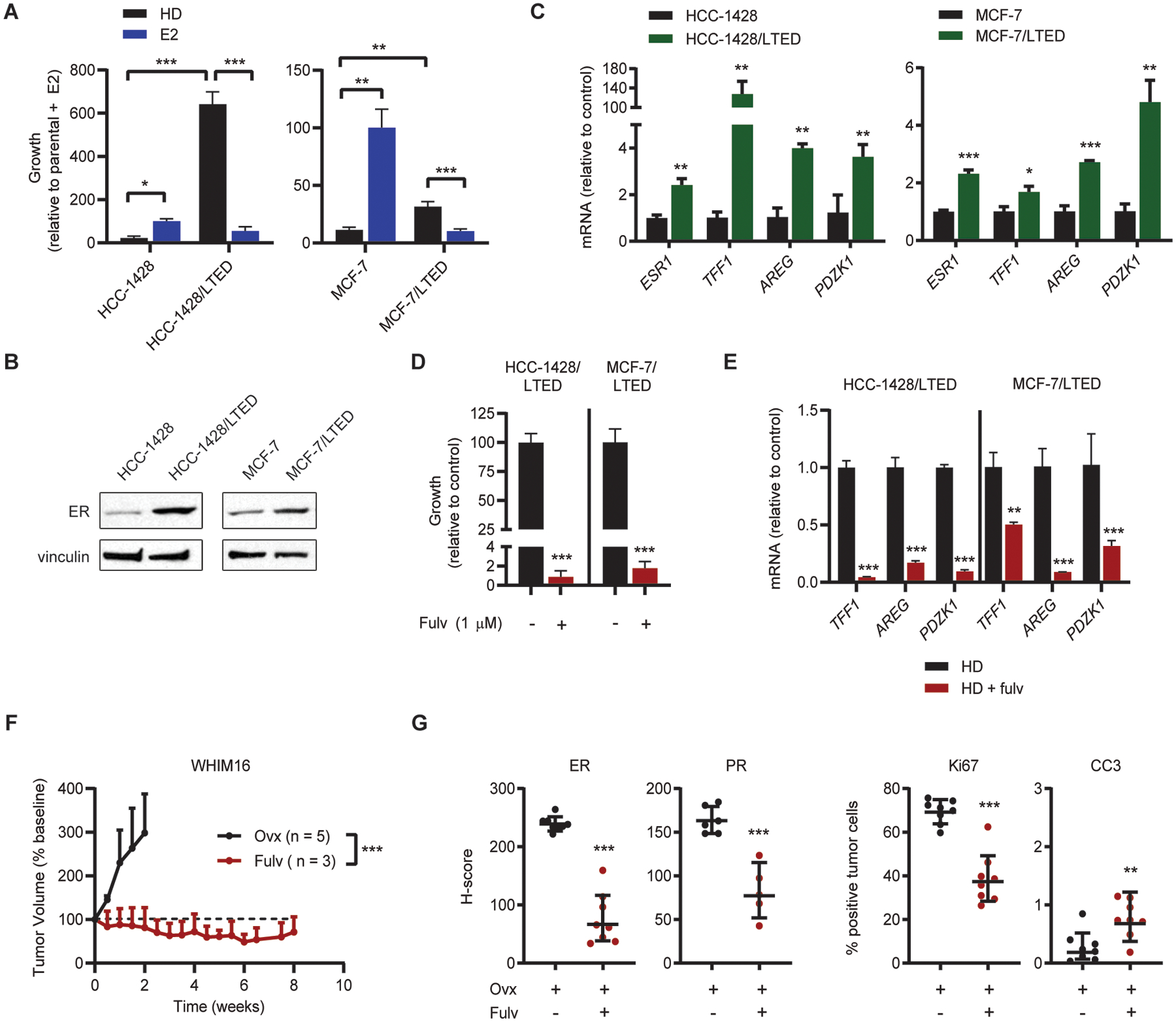
(A) Cells were seeded at low density and treated in triplicate as indicated for 4 wk. Colonies were fixed and stained with crystal violet, and colony area was quantified. Parental cells were hormone-deprived (HD) for 3 d prior to seeding. (B) Lysates from parental cells (hormone-deprived for 3 d) and LTED cells were analyzed by immunoblot. (C) RNA isolated from parental cells (hormone-deprived for 3 d) and LTED cells was analyzed by RT-qPCR. Values for indicated transcripts were normalized to ACTB expression. (D) LTED cells were seeded at low density and treated in triplicate ± fulvestrant (fulv) as indicated for 4 wk. Colony area was analyzed as in (A). (E) LTED cells were treated ± 1 μM fulv for 3 d. RNA was isolated and analyzed by RT-qPCR as in (B). (F) Ovx mice bearing WHIM16 tumors were randomized to treatment as indicated. (G) Tumors were harvested from mice treated as in (F) after 8 d of treatment. Expression of ER, Ki67, cleaved caspase 3 (CC3) and progesterone receptor (PR) were analyzed by IHC. *p≤0.05, **p≤0.005, ***p≤0.0005 by two-tailed t-test (D, G), Bonferroni multiple comparison-adjusted post-hoc test (C, E), or linear mixed modeling (F). Data are shown as mean ± SD.