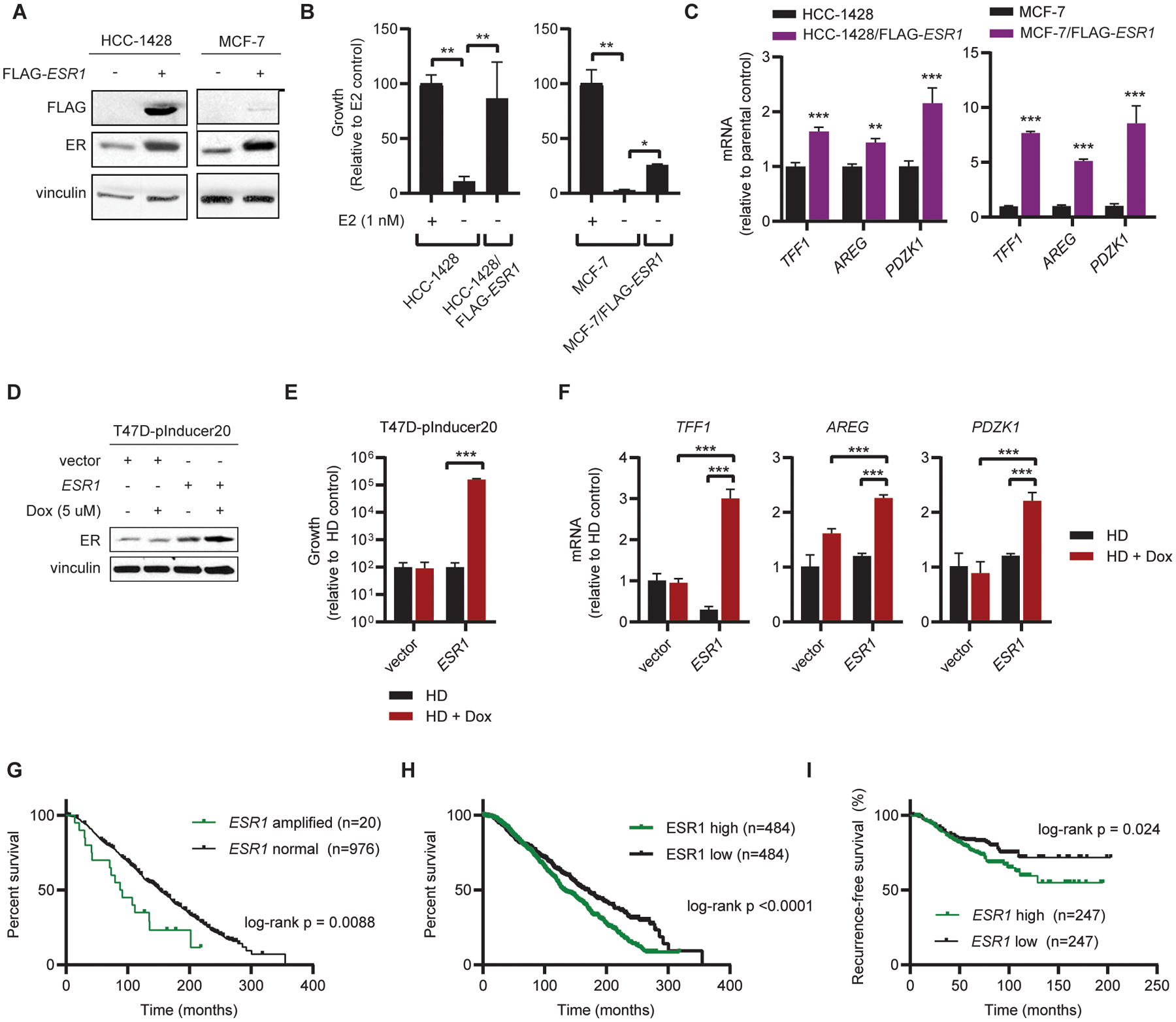
Figure 2. Overexpression of ER induces estrogen-independent ER signaling and resistance to estrogen deprivation.

(A) MCF-7 and HCC-1428 cells stably expressing FLAG-ESR1 were generated, and transgene expression was confirmed by immunoblot. Parental cells were hormone-deprived for 3 d prior to collection of lysates. (B) Parental and FLAG-ESR1 cells were seeded at low density and treated as indicated for 4 wk. Parental cells were hormone-deprived for 3 d prior to seeding. Colonies were fixed and stained, and relative colony area was quantified. (C) RNA was isolated from ER-overexpressing cells and parental controls. Expression of indicated transcripts was analyzed by RT-qPCR, normalized to expression of ACTB. Parental cells were hormone-deprived for 3 d prior to RNA isolation. (D) Protein was harvested from T47D cells stably expressing the pInducer20 vector with either doxycycline-inducible ESR1 or empty vector control. Cells were hormone-deprived for 3 d and treated with doxycycline (dox) as indicated for 2 d. ER expression was analyzed by immunoblot. (E) T47D/pInducer20 cells were hormone-deprived for 3 d and seeded at low density. Cells were treated as indicated in triplicate for 4 wk, and colony area was quantified as in (B). (F) T47D/pInducer20 cells were hormone-deprived for 5 d and treated ± 5 μM doxycycline on Day 3 of hormone deprivation. RNA was harvested and analyzed as in (C). (G) Overall survival analysis of patients with ESR1-amplified (n=20) and -non-amplified (n=976) ER+/HER2- breast cancer. Analysis is limited to patients with ER+ status confirmed by IHC, HER2- status measured by SNP6 array, and receipt of endocrine therapy in ref. (17). (H) Overall survival analysis of patients with high (n=484) vs. low (n=484) ESR1 mRNA expression from dataset in (G). (I) Recurrence-free survival analysis of 494 patients with ESR1-high (n= 247) vs. ESR1-low (n=247) ER+/HER2- breast cancer described in ref. (19). Data were stratified by median ESR1 mRNA expression. Data in (B-F) are shown as mean ± SD. *p≤0.05, **p≤0.005, ***p≤0.0005 by two-tailed t-test (C) or Bonferroni multiple comparison-adjusted post-hoc test (B, E, F).
