
Figure 4. Therapeutic response to E2 is accompanied by hyperactivation of ER transcriptional activity.
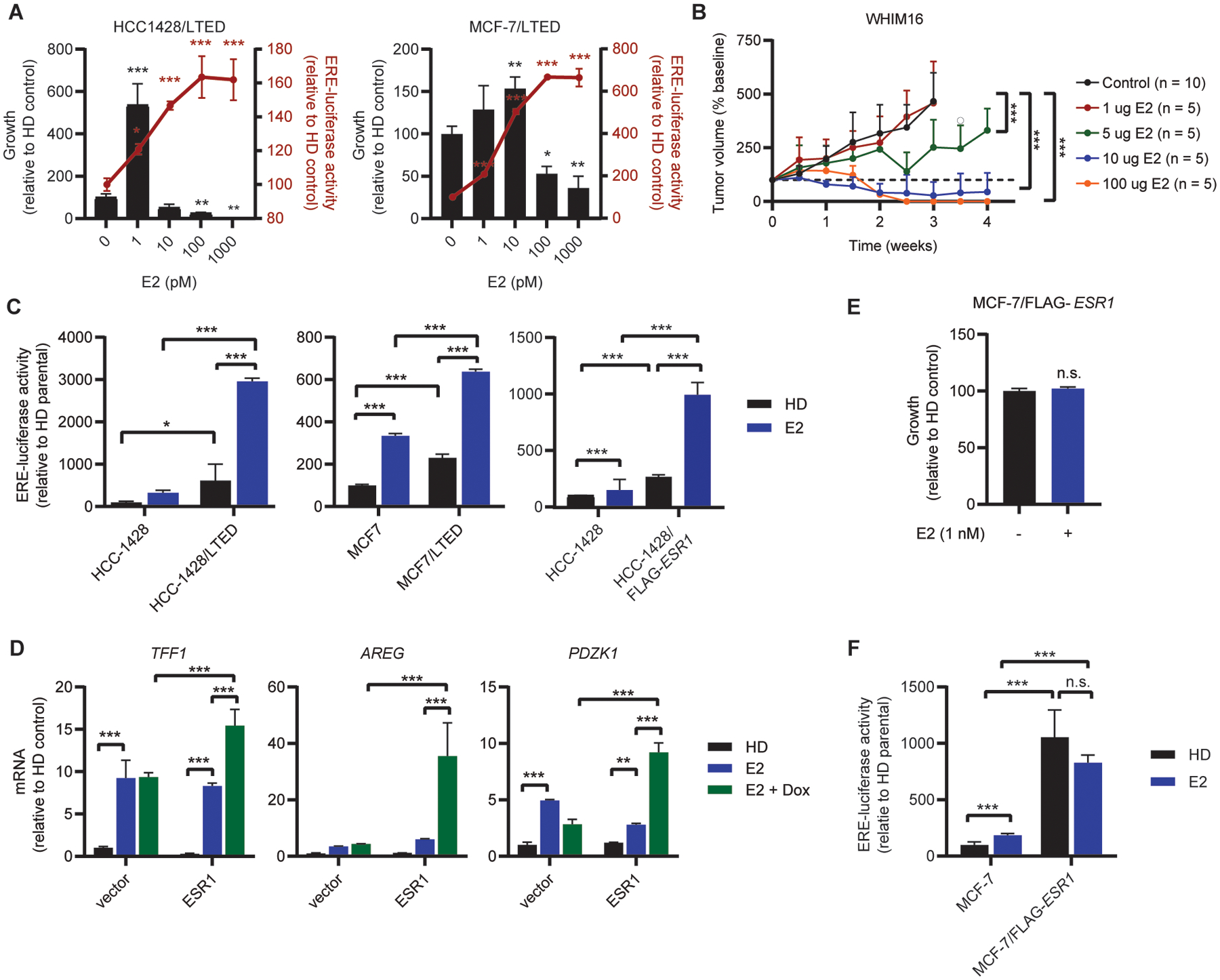
(A) Cells were seeded at low density and treated with E2 as indicated for 4 wk, then fixed and stained with crystal violet. Colony area was quantified using ImageJ (left y-axis). To measure ER activity, cells were transfected with ERE-driven firefly luciferase and CMV-Renilla. One day following transfection, cells were treated with E2 as indicated for 24 h, and luciferase activities were measured (right y-axis). (B) Ovx mice bearing WHIM16 tumors were randomized to treatment with E2 (p.o., BID) at the indicated doses. Tumor volumes were measured twice weekly. Data are shown as mean + SD. (C) HCC-1428 and MCF-7 parental, LTED, and FLAG-ESR1 cells were transfected with luciferase vectors and analyzed as in (A). Parental cells were hormone-deprived for 3 d prior to transfection. (D) RNA was isolated from T47D-pInducer20 cells treated ± 5 μM doxycycline for 2 wk, and ± 1 nM E2 for 24 h. Expression of indicated transcripts was analyzed by RT-qPCR and normalized to ACTB. (E) MCF-7/FLAG-ESR1 cells were seeded at low density, treated with E2 as indicated for 4 wk, and colony area analyzed as in (A). (F) MCF-7/FLAG-ESR1 and MCF-7 parental cells were transfected with luciferase vectors and analyzed as in (A). Parental cells were hormone-deprived for 3 d prior to transfection. Data are shown as mean of 3 (A/C/D/E) or 6 (F) replicates ± SD. *p≤0.05, **p≤0.005, ***p≤0.0005 by Bonferroni multiple comparison-adjusted post-hoc test (A/C/D/F), two-tailed t-test (E) or linear mixed modeling (B).