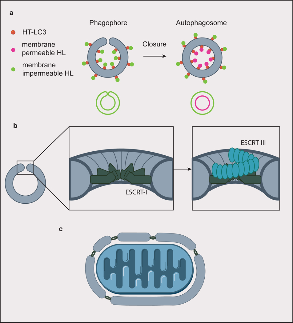
Fig. 4. Autophagosome closure.
(a) A screen for factors involved in autophagosome closure identified ESCRT-I protein VPS37A by using membrane permeable/impermeable Halo ligand (HL) dyes to selectively label Halo tagged (HT) LC3. Hits were identified from cells whose phagophores only stained with the impermeable dye, indicating failure to close the autophagosomes. Closed autophagosomes stain with both the membrane permeable and impermeable dyes. (b) ESCRT-I forms a dodecameric ring assembly at the rim of the closing autophagosome. In canonical ESCRT-mediated membrane scission, subsequent helical assembly of ESCRT-III constricts the membrane rim. (c) For larger cargoes, ESCRTs could be involved in piecing together membranes to form a single, large phagophore.

