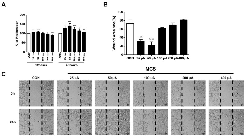
Figure 1.
(A) Effects of MCS on the proliferation of HFDPC cells. To induce cell proliferation, MCS was applied to the HFDPC for 1 h, and the cell was subsequently cultured for 12 and 48 h and incubated further with WST-1 reagent for additional 1 h. The values shown represent the mean ± SD of triplicate measurements of separate experiments. Values are shown as percentages of the control. (B) Relative wound area rate was calculated as the ratio of the remaining wound area at 24 h and the original area at 0 h (C) In vitro scratch assay. Black dotted lines indicate the wound borders at the beginning of the assay and were recorded at 0 and 24 h post-scratching. HFDPC cells were treated with various levels of MCS or left untreated. ** p < 0.01, *** p < 0.001, **** p < 0.0001 vs. control group.

