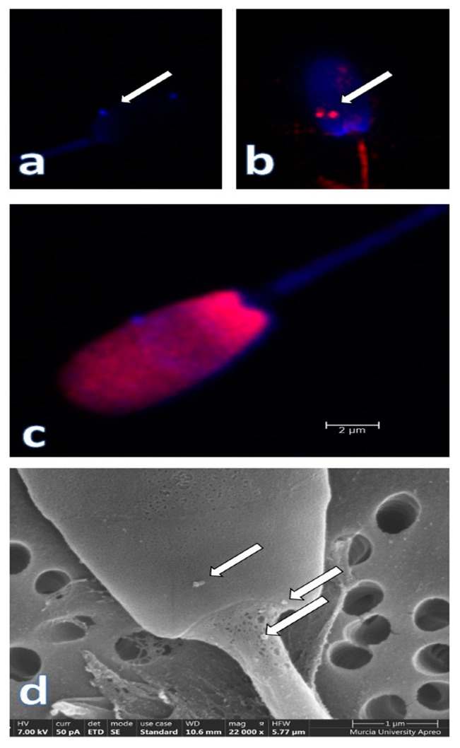
Figure 4.
Seminal plasma extracellular vesicles (EVs) attached to the plasmalemma of ejaculated pig spermatozoa (arrows). Confocal microscopy microphotographs show the CD63 (a) (blue, Alexia fluor 405) and CD9 (b) (red, phycoerythrin, arrows) immunostained EVs. In (c), a composite image of a spermatozoon stained with Hoechst 33,342 and propidium iodide, while a scanning electron microscopy (SEM) micrograph showing some EVs attached to the membrane in the neck and head sperm domains is shown in (d). Courtesy of our graduates L Padilla and I Barranco.

