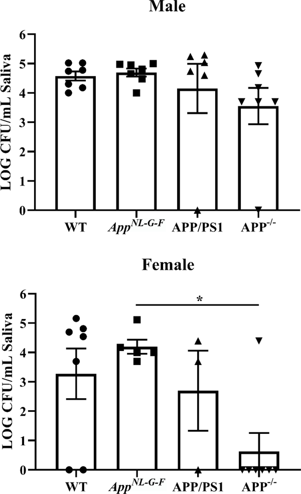
Fig. 6. APP−/− mice had altered oral bacterial growth compared to AppNL–G–F mice.
Pilocarpine-stimulated saliva collected from male and female WT, AppNL–G–F, APP/PS1, and APP−/− mice was plated onto blood agar plates. Numbers of colonies were counted at 24 hours and normalized per volume of saliva from each condition. Data are graphed as mean values +/− SEM. Statistical differences were calculated by one-way ANOVA with Tukey post-hoc analysis, *p<0.05 ( n=3–7).

